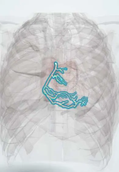
Right coronary artery !
The right coronary artery circles the right side of the heart; it feeds the right ventricle, the underside of the heart and usually its pacemaker.
Opens in the Durgical app, no sign-up needed.
What it is
The right coronary artery, running in the right groove around to the back of the heart; in most people it gives the posterior descending artery.
What it does
Feeds the right ventricle, the bottom of the left ventricle and, in most people, the SA and AV nodes (so its blockage can slow the heart).
Key facts
- Origin
- Right aortic sinus (above right coronary cusp); runs in right coronary groove
- Branches
- Conus (infundibular) branch, usually first (own ostium in some hearts); SA nodal (~60%); right marginal (acute marginal); AV nodal (~80%); posterior descending in right-dominant hearts (~70%, varies); posterolateral branches to inferior LV wall
- Supplies
- Right atrium, right ventricle, inferior LV wall, posterior 1/3 of septum, usually SA and AV nodes
Relations
- Course
- From the right aortic sinus it passes forward between the pulmonary trunk and the right auricle, descends in the right coronary groove to the acute margin, then follows the inferior surface to the crux
- Above
- Right auricle and right atrium
- Below
- Right ventricle, along whose acute margin the right (acute) marginal branch runs
- Superficial
- Epicardial fat and the small cardiac vein, which shares the right coronary groove
How to find it
After opening the pericardium it is followed in the right part of the coronary groove, between the right atrium and right ventricle, embedded in fat; it then turns onto the diaphragmatic surface, where its distal part and the posterior descending artery are reached for grafting by lifting the apex.
On imaging
On coronary angiography it looks like a 'C' in the left anterior oblique view and an 'L' in the right anterior oblique view; it is divided into proximal, mid and distal segments, ending in the posterior descending and posterolateral branches. CT coronary angiography shows dominance and anomalous origins.
Common problems
- Coronary artery disease: fatty plaque narrows them, causing chest pain on exertion (angina).
- Heart attack: a plaque breaks open and a clot suddenly blocks the artery.
How it is diagnosed
ECG and troponin for a heart attack; stress test, CT coronary angiography or a catheter angiogram to see narrowings.
Treatment
- Medicines (aspirin, statins, blood-pressure control) and stopping smoking.
- Angioplasty and stent through a catheter.
- Bypass surgery (CABG) for several or complex blockages.
Clinical notes
- AV block in an inferior MI is usually at the AV node, with a narrow-QRS escape rhythm, and often resolves; in an anterior MI, block is lower and carries a worse outlook
- A proximal occlusion, before the right ventricular branches, adds right ventricular infarction; a distal occlusion spares the right ventricle
- Bypass grafts usually go to its distal part or to the posterior descending artery
- Its conus branch meets the conus branch of the LAD on the front of the right ventricular outflow tract (Vieussens' arterial ring), a collateral route that can open when either artery is blocked
Anatomical variants
- The conus branch often has its own opening in the right aortic sinus (the 'third coronary artery')
- Anomalous origin from the left aortic sinus with a course between the aorta and the pulmonary trunk: linked to ischemia and sudden death in young athletes
Development
Coronary arteries grow from a vessel plexus laid down by the epicardium, which then links up with the aortic sinuses; a faulty link-up explains the anomalous origins listed under variants.
Related conditions
- Atherosclerosis and dyslipidaemia
- Vasospastic (Prinzmetal) angina
- Kawasaki disease
- Multisystem inflammatory syndrome in children (MIS-C)
- Myocardial Infarction (Heart Attack)
- Stable angina
Test yourself
Think of the answer, then tap the question to see it.
Which ECG pattern points to an inferior myocardial infarction from right coronary artery occlusion?
ST elevation in II, III and aVF, higher in III than in II.
Why can nitrates and diuretics drop the blood pressure sharply in right ventricular infarction?
The heart depends on filling; the picture is low blood pressure with clear lungs and raised jugular venous pressure.
Why do bradycardia and AV block occur in an inferior myocardial infarction?
Ischemia of the SA and AV nodal branches of the right coronary artery, plus vagal reflexes (Bezold–Jarisch reflex).
Which nodal and septal branches does the right coronary artery usually give?
SA nodal (about 60%), AV nodal (about 80%) and, in right-dominant hearts, the posterior descending artery to the posterior 1/3 of the septum.
What shape does the right coronary artery have on angiography?
A «C» in the left anterior oblique view and an «L» in the right anterior oblique view.
Educational information, not medical advice. For any symptom, see a health professional.
Its names
- Official Latin (TA2)
- arteria coronaria dextra; arteria interventricularis inferior TA2 4131
- Official Spanish (DeCS)
- Arteria coronaria derecha DeCS D003331
- English (TA2)
- Right coronary artery
- Also called
- posterior interventricular artery · inferior intervenricular branch of right coronary artery · posterior interventricular branch of right coronary artery · RCA · PDA · arteria coronaria cordis dextra
Related
- Posterior descending artery (PDA)
- Right atrium
- Right ventricle
- Sinoatrial (SA) node
- Atrioventricular (AV) node
- Left ventricle
- Left ventricle papillary muscles
Sources: Terminologia Anatomica (TA2); DeCS; Anatomy, Thorax, Heart; Anatomy, Thorax, Sinoatrial Nodal Artery; 19.1 Heart Anatomy (OpenStax (Rice University), 2022) · adapted, CC BY 4.0; Tratado de anatomía humana (9.ª ed.), tomo II, Libro IV Angiología, sección 1, cap. I Corazón (arts. VI–IX) y cap. II Pericardio.
Spotted a mistake?We are always improving for the community. Tell us what is wrong and our team will review it.Report a mistake
Representative model, for educational purposes only.