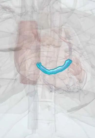
Coronary sinus !
The coronary sinus is a large vein in the groove at the back of the heart; it collects the heart's own used blood and empties it into the right atrium.
Opens in the Durgical app, no sign-up needed.
What it is
A wide vein in the groove on the back of the heart that collects blood from the cardiac veins.
What it does
Empties the heart's own used blood into the right atrium. Pacemaker leads for resynchronization travel through it to reach the left ventricle.
Key facts
- Drains
- Most of heart's venous blood: great, middle and small cardiac veins, posterior LV vein, oblique vein of left atrium
- Into
- Right atrium, between IVC opening and tricuspid orifice (valve of coronary sinus); lies in posterior coronary groove
Relations
- Course
- Lies in the posterior part of the coronary (atrioventricular) groove on the diaphragmatic surface, running from left to right between the left atrium and the left ventricle to the right atrium
- Above
- Left atrium; the oblique vein of the left atrium (vein of Marshall) joins its left end
- Below
- Base of the left ventricle
- Deep
- Mitral annulus, close by, and the circumflex artery, which often passes between the two
How to find it
On the back of the heart it lies in the coronary groove between the left atrium and left ventricle. Its opening in the right atrium sits between the inferior vena cava opening and the tricuspid orifice; catheters reach it from the right atrium, entered from a vein of the neck, chest or groin.
On imaging
On echocardiography it appears as a small round channel in the posterior atrioventricular groove, behind the mitral annulus, in the parasternal long-axis view. Coronary sinus venography or CT venography maps its tributaries before a resynchronization lead is placed.
Common problems
- Rarely a problem on its own.
Clinical notes
- Placing the left ventricular lead can be hindered by a prominent Thebesian valve, sharp bends or few suitable tributaries; stimulation of the left phrenic nerve is a known problem with lateral leads
- Electrophysiology catheters in the coronary sinus record left atrial and left ventricular signals and help map left-sided accessory pathways
- Unroofed coronary sinus: a rare atrial septal defect in which the wall between the sinus and the left atrium is missing
Anatomical variants
- Persistent left superior vena cava draining into the coronary sinus, the commonest thoracic venous anomaly; on echocardiography the sinus looks dilated
- The Thebesian valve and the valve of Vieussens vary in size and shape and can obstruct catheters
Development
Derives from the left horn of the sinus venosus; a persistent left superior vena cava is the left anterior cardinal vein that failed to regress.
Related conditions
Test yourself
Think of the answer, then tap the question to see it.
Where does the coronary sinus open, and what landmark does its opening form?
Into the right atrium between the inferior vena cava opening and the tricuspid orifice, guarded by the Thebesian valve; it forms the base of the triangle of Koch.
How does the left ventricular lead of cardiac resynchronization reach the left ventricle?
From the right atrium into the coronary sinus and on into a lateral or posterolateral tributary.
What is retrograde cardioplegia?
Protective solution run backwards through a balloon catheter in the coronary sinus to reach heart muscle beyond blocked arteries.
Which venous anomaly drains into the coronary sinus and makes it look dilated on echo?
A persistent left superior vena cava, the commonest thoracic venous anomaly.
Educational information, not medical advice. For any symptom, see a health professional.
Its names
- Official Latin (TA2)
- sinus coronarius TA2 4158
- Official Spanish (DeCS)
- Seno coronario DeCS D054326
- English (TA2)
- Coronary sinus
Related
Sources: Terminologia Anatomica (TA2); DeCS; Anatomy, Thorax, Coronary Sinus; 19.1 Heart Anatomy (OpenStax (Rice University), 2022) · adapted, CC BY 4.0; Tratado de anatomía humana (9.ª ed.), tomo II, Libro IV Angiología, sección 1, cap. I Corazón (arts. VI–IX) y cap. II Pericardio.
Spotted a mistake?We are always improving for the community. Tell us what is wrong and our team will review it.Report a mistake
Representative model, for educational purposes only.