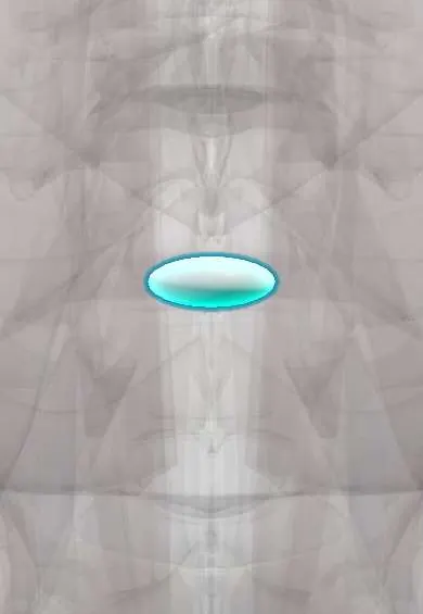
L4–L5 nucleus !
The nucleus pulposus is the soft, gel-like centre of the L4-L5 disc in the lower back; it is the part that bulges out in a disc herniation.
Official name: L4-L5
Opens in the Durgical app, no sign-up needed.
What it is
The soft, gel-like centre of the L4-L5 disc (nucleus pulposus), mostly water held by proteins that attract it.
What it does
Works like a water cushion: it turns pressure from above into outward push on the fibre ring, spreading the load. It is under the most pressure when you bend forward and lift.
Key facts
- Where
- Centre of the L4–L5 disc, slightly toward the back, inside the annulus fibrosus
- Function
- Notochord-derived gel (about 70–90% water, proteoglycans); turns axial load into annulus tension; herniates posterolaterally toward the L5 root
Relations
- In front
- Front of the annulus and the anterior longitudinal ligament, then the common iliac arteries and the start of the inferior vena cava, which an instrument pushed through the front of the disc during discectomy can injure
- Behind
- Posterior longitudinal ligament, then the dural sac; the L5 root in the lateral recess is the one a posterolateral fragment usually compresses
- Lateral
- The L4 root in the foramen, reached by a foraminal or far-lateral fragment
Examination
- Straight leg raise: pain below the knee between about 30° and 70° suggests L5 or S1 root tension; sciatica felt when the healthy leg is raised (crossed test) is less sensitive but more specific
- L5 root: weak big-toe extension and heel walking, numb top of the foot; reflexes are often normal
On imaging
Urgent MRI confirms a central compression. After surgery, contrast MRI separates recurrent herniation (a fragment that does not enhance, with an enhancing rim) from scar (which enhances throughout).
Common problems
- Squeezes out through a tear in the ring (herniation), usually backward and to one side, toward a nerve root.
- Loses water with age, so the disc gets thinner and stiffer.
How it is diagnosed
On MRI a healthy nucleus is bright on T2 images; a worn one turns dark ('black disc').
Treatment
- In a microdiscectomy only the loose piece that has pushed out is removed; most of the disc stays.
Clinical notes
- Microdiscectomy removes the herniated fragment and loose nucleus, not the whole nucleus; what remains can herniate again at the same level
- Degenerative spondylolisthesis is most frequent at L4–L5, typically in women over 50: disc collapse with facet arthritis lets L4 slip forward and narrows the canal
- In anterior lumbar interbody fusion at L4–L5 the aorta, the vena cava and the left common iliac vein must be moved aside; venous tearing is the main vascular risk, and the iliolumbar vein is often tied first
Related conditions
Test yourself
Think of the answer, then tap the question to see it.
What can a large central extrusion of the L4–L5 nucleus cause?
Cauda equina syndrome: compression of the whole cauda equina, a surgical emergency.
Which red flags are taught for cauda equina syndrome from an L4–L5 nucleus herniation?
Saddle numbness, new trouble passing or holding urine, loss of bowel control, sexual dysfunction, sciatica in both legs and worsening weakness.
After surgery on the L4–L5 nucleus, how does contrast MRI separate recurrent herniation from scar?
A recurrent fragment does not enhance, apart from an enhancing rim; scar enhances throughout.
What does a microdiscectomy remove from the L4–L5 nucleus?
Only the herniated fragment and loose nucleus, not the whole nucleus; what remains can herniate again at the same level.
How do a healthy and a worn L4–L5 nucleus look on T2 MRI?
A healthy nucleus is bright; a worn one, which has lost water, turns dark (the «black disc»).
Educational information, not medical advice. For any symptom, see a health professional.
Its names
- Official Latin (TA2)
- nucleus pulposus L4-L5 TA2 1686
- Official Spanish (DeCS)
- Núcleo pulposo L4-L5 DeCS D000070614
- English (TA2)
- L4-L5
Related
Sources: Terminologia Anatomica (TA2); DeCS; Anatomy, Back, Nucleus Pulposus; Anatomy, Back, Intervertebral Discs; 7.3 The Vertebral Column (OpenStax (Rice University), 2022) · adapted, CC BY 4.0.
Spotted a mistake?We are always improving for the community. Tell us what is wrong and our team will review it.Report a mistake
Representative model, for educational purposes only.