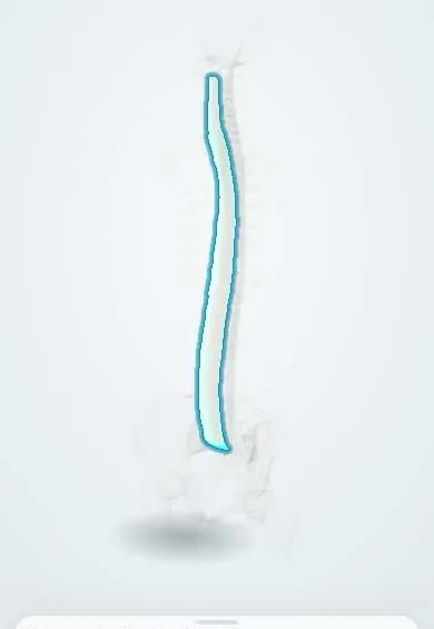
Anterior longitudinal ligament !
The anterior longitudinal ligament is a long, wide band down the front of the vertebrae and discs; it stops the spine bending too far back.
Opens in the Durgical app, no sign-up needed.
What it is
A long, wide band running down the front of all the vertebral bodies and discs, from the skull base to the sacrum.
What it does
Stops the spine from bending too far backward (extension) and reinforces the front of the discs.
Key facts
- Attaches
- Front of vertebral bodies and discs, from basilar occiput and anterior tubercle of atlas to the pelvic surface of the sacrum
- Function
- Limits extension; reinforces the front of the discs; strong and wide
- Tight in
- Extension (bending backward)
Relations
- In front
- Neck: longus colli, prevertebral fascia, pharynx and oesophagus; chest: descending aorta, azygos vein, thoracic duct; abdomen: abdominal aorta, inferior vena cava, diaphragm crura
- Behind
- Front of the vertebral bodies and of the annulus fibrosus of each disc
- Lateral
- The sympathetic trunks along the sides of the bodies; in the lumbar region, the psoas origins and the segmental lumbar arteries crossing the waists of the bodies
How to find it
Not palpable. In the anterior cervical approach it is the shiny band on the front of the vertebral bodies, between the two longus colli muscles, once the prevertebral fascia is opened. In the lumbar spine it lies on the bodies behind the aorta, vena cava and iliac vessels, between the origins of the psoas.
On imaging
MRI shows it as a thin dark line on the front of the bodies; after hyperextension, a gap or high signal, often with prevertebral swelling on the lateral radiograph. CT tells the ossification patterns apart: thick flowing bone in DISH, thin marginal syndesmophytes in ankylosing spondylitis.
Common problems
- Tears in whiplash or other hyperextension injuries.
- Turns to bone in DISH (diffuse idiopathic skeletal hyperostosis) and in ankylosing spondylitis, stiffening the spine.
How it is diagnosed
MRI for tears; X-rays and CT show ossification.
Treatment
- Usually treated through the underlying condition. In ALIF it is opened at one level to reach the disc.
Clinical notes
- Anterior approaches (anterior cervical discectomy, anterior lumbar interbody fusion) cut through it to reach the disc
- Large anterior cervical osteophytes or DISH can press on the oesophagus and cause difficulty swallowing
- An extension teardrop fragment from the lower front corner of a body (often C2) marks an avulsion at its attachment
Related conditions
Test yourself
Think of the answer, then tap the question to see it.
Where does the anterior longitudinal ligament attach?
To the front of the vertebral bodies and discs, from the basilar occiput and anterior tubercle of the atlas down to the pelvic surface of the sacrum.
Which movement does the anterior longitudinal ligament limit?
Extension: it is the main restraint to hyperextension, wider and stronger than the posterior longitudinal ligament.
What can a hyperextension injury cause in an older neck with spondylosis when it tears the anterior longitudinal ligament?
Central cord syndrome, even without any fracture.
What are the features of DISH, which ossifies the anterior longitudinal ligament?
Flowing ossification along the front and sides of four or more adjacent vertebrae, on the right in the thoracic spine (the aorta lies on the left), with disc heights preserved.
Why are fractures dangerous in a spine fused by ankylosing spondylitis or DISH, where the anterior longitudinal ligament has ossified?
Minor trauma can break through all three columns, and these fractures are easily missed.
Educational information, not medical advice. For any symptom, see a health professional.
Its names
- Official Latin (TA2)
- ligamentum longitudinale anterius TA2 1679
- Official Spanish (DeCS)
- Ligamento longitudinal anterior DeCS D017846
- English (TA2)
- Anterior longitudinal ligament
- Also called
- ALL · ligamentum longitudinale commune ventrale
Related
- Posterior longitudinal ligament
- Axis (C2)
- C3 vertebra
- C4 vertebra
- C5 vertebra
- C6 vertebra
- C7 vertebra
- L1 vertebra
Sources: Terminologia Anatomica (TA2); DeCS; Anatomy, Back, Posterior Longitudinal Ligament.
Spotted a mistake?We are always improving for the community. Tell us what is wrong and our team will review it.Report a mistake
Representative model, for educational purposes only.