Linea alba !
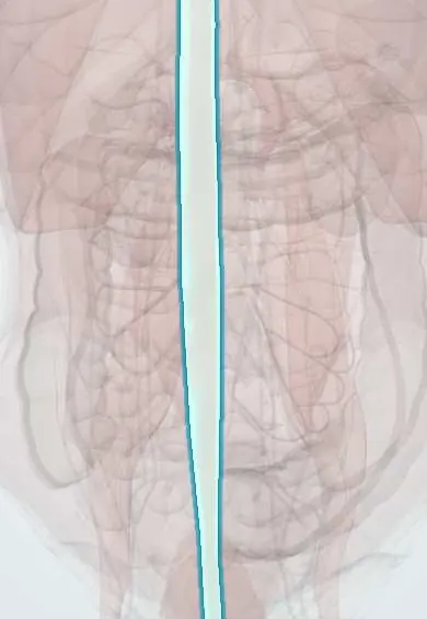
The linea alba is the band of tough tissue running down the middle of the belly, from the breastbone to the pubis, between the two rectus muscles.
Opens in the Durgical app, no sign-up needed.
What it is
A tough midline seam of tendon from the breastbone to the pubis, where the muscle sheets of both sides meet.
What it does
Anchors the abdominal muscles. It has few blood vessels, so it is a common incision line.
Key facts
- Where
- Anterior midline from xiphoid process to pubic symphysis and pubic crest; wider above the umbilicus; contains the umbilical ring
- Attaches
- Interlacing aponeuroses of external oblique, internal oblique and transversus abdominis of both sides
- Function
- Midline anchor of the flat abdominal muscles; relatively avascular plane used for midline incisions; site of epigastric hernias
Relations
- In front
- Subcutaneous fat and skin; the umbilicus, usually at about the L3–L4 level
- Behind
- Transversalis fascia, extraperitoneal fat and peritoneum; above the umbilicus the falciform ligament with the round ligament of the liver, below it the median umbilical ligament (urachus remnant)
- Lateral
- The rectus sheath and rectus abdominis of each side
- Above
- The xiphoid process
- Below
- The pubic symphysis and pubic crest; the pyramidalis inserts into its lower part
How to find it
In a lean person it shows as a shallow vertical groove between the two recti from the xiphoid to the umbilicus; below the umbilicus it is barely visible. In pregnancy the skin over it often darkens as the linea nigra.
Examination
- Head-and-shoulder raise (curl-up): a midline ridge or a gap between the recti that the examiner's fingers can sink into shows diastasis recti; the gap is measured in finger-breadths or by ultrasound
- Cough impulse: a midline bulge in the epigastrium or at the umbilicus that pushes against the hand on coughing suggests a hernia through the linea alba
On imaging
A linear ultrasound probe shows it as a thin bright band between the two rectus muscles; CT shows it on axial slices. Tell diastasis recti (a widened gap that bulges on straining, with no fascial defect) from an epigastric or umbilical hernia, where fat or bowel passes through a hole. Dynamic ultrasound with Valsalva, or CT, is used when the examination is unclear.
Common problems
- Epigastric and umbilical hernias.
- Widening in diastasis recti.
- Incisional hernia after midline surgery.
How it is diagnosed
Examination, often while coughing or standing; ultrasound or CT if unclear.
Treatment
- Hernias that cause symptoms are repaired, usually with a mesh.
Clinical notes
- Epigastric hernia: small defects in the crossing fibres between the xiphoid and the umbilicus, often holding only extraperitoneal fat; there may be several
- Infant umbilical hernia through the umbilical ring usually closes on its own in early childhood; the adult paraumbilical type does not and is more prone to trapping bowel
- Incisional hernia after midline laparotomy; how the linea alba is closed (suture length and small bites) affects the risk
- Diastasis recti: common after pregnancy and in newborns; it causes a midline bulge on straining but no trapping of bowel
Development
The umbilical ring is where the umbilical cord structures passed. The midgut normally herniates into the cord and returns by about week 10–12; failure leaves an omphalocele (covered by a sac), whereas gastroschisis is a defect beside the cord, usually on the right, with no sac.
Related conditions
Test yourself
Think of the answer, then tap the question to see it.
Why are epigastric hernias and diastasis recti mostly above the umbilicus?
The linea alba is broad above the umbilicus and almost a thin line below it, so defects in its crossing fibres and widening occur mainly between xiphoid and umbilicus.
What forms the linea alba?
The interlacing aponeuroses of the external oblique, internal oblique and transversus abdominis of both sides, running from the xiphoid process to the pubic symphysis and pubic crest.
What is the main late complication of a midline laparotomy through the linea alba?
Incisional hernia: the quick, nearly bloodless incision divides no muscle or nerve but heals as scar; closure technique (suture length, small bites) affects the risk.
How does diastasis recti differ from a hernia of the linea alba?
Diastasis recti is a widened, thinned linea alba with no defect and no hernia sac; it bulges on straining but does not trap bowel.
Educational information, not medical advice. For any symptom, see a health professional.
Its names
- Official Latin (TA2)
- linea alba TA2 2377
- Official Spanish (DeCS)
- Línea alba
- English (TA2)
- Linea alba
Related
Sources: Terminologia Anatomica (TA2); Anatomy, Abdomen and Pelvis: Abdominal Wall; Anatomy, Abdomen and Pelvis, Rectus Sheath.
Spotted a mistake?We are always improving for the community. Tell us what is wrong and our team will review it.Report a mistake
Representative model, for educational purposes only.