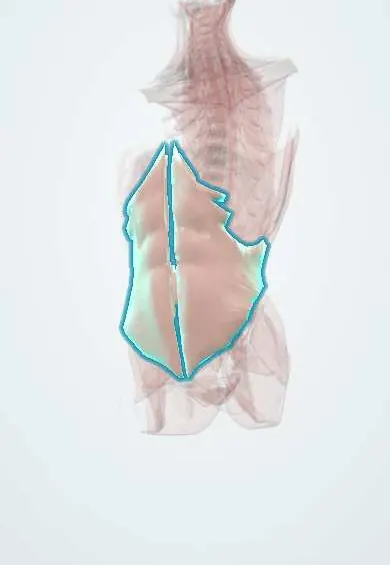
Internal oblique !
The internal oblique is the middle layer of the side abdominal wall; it bends and turns the trunk and helps stiffen the spine.
Official name: Internal abdominal oblique muscle
Opens in the Durgical app, no sign-up needed.
What it is
The middle layer of the side abdominal wall, with fibres running up and forward.
What it does
Bends and turns the trunk to the same side and helps the transversus stiffen the spine.
Key facts
- Origin
- Thoracolumbar fascia, anterior two-thirds of iliac crest, lateral third of inguinal ligament (some texts: lateral two-thirds)
- Insertion
- Inferior borders of ribs 10–12, linea alba, pubis via conjoint tendon
- Action
- Flexes and rotates trunk to the same side; compresses abdomen
- Nerve
- Thoracoabdominal nerves (T7–T11), subcostal (T12), iliohypogastric and ilioinguinal (L1)
- Blood supply
- Deep circumflex iliac, superior and inferior epigastric, lower posterior intercostal arteries
Relations
- Superficial
- External oblique, with fibres crossing at roughly a right angle
- Deep
- Transversus abdominis; the lower thoracic, subcostal, iliohypogastric and ilioinguinal nerves and their vessels run in the plane between them
- Medial
- Rectus sheath, which its aponeurosis helps form on both sides of the rectus abdominis above the arcuate line
- Below
- Arches over the inguinal canal as its roof; the ilioinguinal nerve pierces it near the ASIS and runs on in the canal
- Behind
- Arises from the thoracolumbar fascia; it forms the floor of the inferior lumbar triangle (of Petit) and a border of the superior lumbar triangle (of Grynfeltt)
How to find it
It cannot be felt apart from the external oblique. On ultrasound of the flank it is the middle and usually the thickest of the three muscle layers, between the external oblique and the transversus abdominis.
Examination
- Cremasteric reflex: stroking the upper inner thigh makes the testis rise on that side; an absent reflex points to an L1–L2 or genitofemoral nerve lesion and is typical of testicular torsion
On imaging
On flank ultrasound it is the middle layer, usually the thickest, between the external oblique and transversus abdominis; near the ASIS the ilioinguinal and iliohypogastric nerves are found between it and transversus for a block. In the groin, ultrasound with straining or CT shows inguinal hernias; a direct hernia bulges medial to the inferior epigastric vessels, through a weak conjoint area.
Common problems
- Delayed or weak activation is seen in some people with chronic low back pain.
How it is diagnosed
Exam; ultrasound can show its activation.
Treatment
- Core stability exercise programmes.
Clinical notes
- The ilioinguinal and iliohypogastric nerves run on or through it near the ASIS and can be caught in hernia repair sutures or mesh, a cause of chronic groin pain
- A weak conjoint tendon favours direct inguinal hernias through the inguinal (Hesselbach) triangle
- The transversus abdominis plane (TAP) block places local anaesthetic in the plane just deep to it, where the abdominal wall nerves run
Related conditions
Test yourself
Think of the answer, then tap the question to see it.
What do the lowest fibres of the internal oblique form in the inguinal region?
The roof of the inguinal canal, then, with the transversus abdominis, the conjoint tendon on the posterior wall.
Which muscle derives from the internal oblique, and what supplies it?
The cremaster, wrapping the spermatic cord; supplied by the genital branch of the genitofemoral nerve (L1–L2).
Which way does the internal oblique rotate the trunk?
To the same side; it also flexes the trunk and compresses the abdomen.
What does an absent cremasteric reflex suggest?
A lesion of L1–L2 or the genitofemoral nerve; it is typical of testicular torsion.
Educational information, not medical advice. For any symptom, see a health professional.
Its names
- Official Latin (TA2)
- musculus obliquus internus abdominis TA2 2373
- Official Spanish (DeCS)
- Músculo oblicuo interno del abdomen DeCS D000009
- English (TA2)
- Internal abdominal oblique muscle
- Also called
- internal oblique muscle
Related
- Transversus abdominis
- External oblique
- Intercostal nerves
- Iliohypogastric and ilioinguinal nerves
- Hip bone
Sources: Terminologia Anatomica (TA2); DeCS; Anatomy, Anterolateral Abdominal Wall Muscles.
Spotted a mistake?We are always improving for the community. Tell us what is wrong and our team will review it.Report a mistake
Representative model, for educational purposes only.