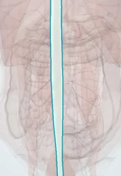
Incisional (ventral) hernia !
An incisional hernia is a bulge through a weak area in an old surgical scar on the belly, most often after a midline abdominal operation.
Updated 2026-10-29
Structures involved
Tap one to see what it is and open it in 3D.
- Linea alba
- Rectus sheath
- Rectus abdominis
- Transversalis fascia
- External oblique
- Internal oblique
- Transversus abdominis
- Peritoneum
- Greater omentum
- Epigastric vessels
In the clinical exam
Why it happens
- What delays healing: infection, diabetes, nutrition, glucocorticoids, poor blood flow
- Healing of skin wounds: first and second intention
- Abnormal repair: chronic wounds, hypertrophic scar, keloid, contracture
- Collagen synthesis
- Wound healing: phases and scar
What it is
A hernia through a weak area in a previous surgical scar of the abdominal wall, most often after a midline laparotomy. It is a type of ventral hernia.
Who gets it
One of the most common long-term complications of open abdominal surgery. Rates vary widely between studies and rise with wound infection, obesity and emergency operations.
Causes
- Failure of the fascial closure after laparotomy
- Surgical site infection or wound dehiscence
- Trocar-site hernia after laparoscopy (especially at the umbilicus)
- Stoma site (parastomal hernia)
Risk factors
- Surgical site infection
- Obesity, smoking and diabetes
- Emergency surgery and midline incisions
- Chronic cough (COPD), constipation, ascites, pregnancy after surgery
- Malnutrition, corticosteroids and immunosuppression
- Abdominal aortic aneurysm and connective tissue disorders
- Closure technique (large bites, rapidly absorbable suture)
Pathophysiology
The fascial edges separate in the weeks after surgery, often before the scar has gained strength. Peritoneum, omentum or bowel then pushes through the gap. Over time the hernia can enlarge until much of the bowel lives outside the abdominal cavity (loss of domain).
Classification
- Ventral hernias: primary (umbilical, epigastric, Spigelian) vs incisional
- European Hernia Society: midline (M1 to M5) or lateral (L1 to L4) location and defect width (W1 to W3)
- Reducible, incarcerated or strangulated; with or without loss of domain
Symptoms and signs
- Bulge in or near a surgical scar, larger when standing or straining
- Discomfort, pulling or a dragging pain
- Skin changes or ulceration over large hernias
- Many are asymptomatic
Diagnosis
- Clinical
- Palpation of the scar standing and lying, with Valsalva; defect size, reducibility, skin quality and previous operations are recorded.
- Tests
- Preoperative tests depend on the patient: glucose control, nutrition and smoking status. Blood count, lactate and electrolytes if strangulation is suspected.
- Imaging
- CT defines the defect size, contents, other hidden defects and loss of domain, and guides the choice of repair. Ultrasound can confirm small hernias.
Differential diagnosis
- Diastasis of the rectus muscles
- Primary umbilical or epigastric hernia
- Seroma, haematoma or abscess in the wound
- Abdominal wall tumour (e.g., desmoid, lipoma)
- Parastomal hernia or prolapse
Goal of treatment
Close the defect with a durable repair that restores abdominal wall function, while optimising the patient beforehand to reduce complications and recurrence.
Treatment
- Follow-up
- Watchful waiting is an option for small, asymptomatic hernias or patients with high surgical risk, with clear warning signs explained.
- Lifestyle
- Prehabilitation before elective repair: weight loss, stopping smoking, glucose control and improved nutrition.
- Surgical
- Open repair with mesh, often placed behind the rectus muscles (retromuscular, Rives-Stoppa type); suture repair alone has high recurrence.
- Surgical
- Laparoscopic or robotic repair with intraperitoneal or extraperitoneal mesh for suitable defects.
- Surgical
- Component separation (anterior or transversus abdominis release) for large defects or loss of domain.
- Acute
- Emergency surgery for incarceration or strangulation, with bowel resection if needed; mesh use in contaminated fields is individualised.
Complications
- Incarceration, strangulation and bowel obstruction
- Recurrence after repair
- Seroma, wound infection and mesh infection
- Enterocutaneous fistula
- Loss of domain and respiratory problems after reduction of very large hernias
Prognosis
Incisional hernias tend to grow. Elective repair with mesh in an optimised patient has the best results; recurrence rises with each new repair and with emergency surgery.
Prevention
- Small-bites closure of midline incisions with slowly absorbable suture, keeping a suture length at least four times the wound length
- Prevention of surgical site infection
- Prophylactic mesh in selected high-risk patients
- Weight control and stopping smoking
Test yourself
Think of the answer, then tap the question to see it.
What is the strongest modifiable risk factor for incisional hernia after laparotomy?
Surgical site infection; obesity and smoking are other important modifiable factors.
Which closure technique for midline laparotomy lowers the rate of incisional hernia?
Small-bites closure with a slowly absorbable continuous suture, with a suture-to-wound length ratio of at least 4:1.
Why is mesh generally used in incisional hernia repair?
Suture repair alone has a high recurrence rate; mesh reinforcement lowers it.
What does loss of domain mean in a large incisional hernia?
A large part of the abdominal contents lives in the hernia sac, so returning it can raise abdominal pressure and impair breathing.
Related conditions
Educational information, not medical advice. For any symptom, see a health professional.
Sources: Ventral Hernia (StatPearls (NCBI Bookshelf), 2023).
Spotted a mistake?We are always improving for the community. Tell us what is wrong and our team will review it.Report a mistake
Representative model, for educational purposes only.