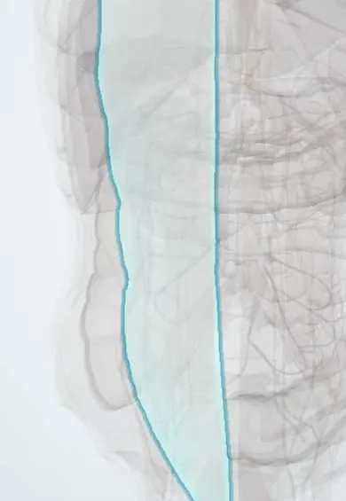
Rectus sheath !
The rectus sheath is the tough fibrous casing around the rectus abdominis, the long vertical muscle at the front of the belly.
Opens in the Durgical app, no sign-up needed.
What it is
The tough envelope around each rectus muscle, made of the tendon sheets of the three flat muscles.
What it does
Holds the rectus in place; surgeons close it carefully after a midline incision to prevent a hernia.
Key facts
- Where
- Aponeurotic sheath around rectus abdominis and pyramidalis, from xiphoid/costal cartilages 5–7 to pubis
- Attaches
- Above arcuate line: front = external + internal oblique, back = internal oblique + transversus; below it all pass in front, transversalis fascia behind
- Function
- Holds rectus in place; contains superior and inferior epigastric vessels and T7–T12 ventral rami; fuses medially as the linea alba
Relations
- In front
- Above the arcuate line: external oblique aponeurosis and the front lamina of the internal oblique aponeurosis; below it: all three flat-muscle aponeuroses; then subcutaneous tissue and skin
- Behind
- Above the costal margin: costal cartilages 5–7 directly (no posterior layer); down to the arcuate line: back lamina of the internal oblique plus the transversus abdominis aponeurosis; below it: transversalis fascia, extraperitoneal fat and peritoneum
- Medial
- The linea alba, where the sheaths of both sides interlace
- Lateral
- The linea semilunaris, where the aponeuroses of external oblique, internal oblique and transversus abdominis meet to split around the muscle
- Contents
- Rectus abdominis and pyramidalis; superior and inferior epigastric vessels; the ends of the lower intercostal nerves and the subcostal nerve, which enter from the side
How to find it
In a lean, muscular person lying on the back who lifts the head and shoulders, the lateral edge of the sheath shows as a curved groove from the tip of the ninth costal cartilage down to the pubic tubercle. The arcuate line can't be felt; at laparoscopy it is seen from inside as the curved lower edge of the posterior layer.
Examination
- Carnett sign: pain that stays the same or worsens when the abdominal muscles are tensed (head raise) points to a source in the wall, such as a sheath hematoma or nerve entrapment, rather than inside the abdomen
- Fothergill sign: a wall mass that stays palpable and does not cross the midline when the rectus abdominis contracts suggests a rectus sheath hematoma
On imaging
Ultrasound shows the rectus abdominis between the bright anterior and posterior layers; it guides the rectus sheath block. On CT a sheath hematoma is a spindle-shaped mass within the muscle; below the arcuate line it is often larger and can track down toward the pelvis.
Common problems
- Hernias: a weak spot lets fat or bowel bulge through (groin, belly button, old incisions).
- Muscle strains.
How it is diagnosed
Examination, often while coughing or standing; ultrasound or CT if unclear.
Treatment
- Hernias that cause symptoms are repaired, usually with a mesh.
Clinical notes
- Rectus sheath hematoma: bleeding from an epigastric artery after violent coughing, trauma or anticoagulation; a painful, tender wall mass
- Rectus sheath block: local anaesthetic placed between the rectus abdominis and the posterior layer numbs the midline skin for umbilical and midline incisions; the TAP block covers the lateral wall instead
- Spigelian hernia: through the aponeurosis along the linea semilunaris, usually near or below the arcuate line; it often lies under the external oblique aponeurosis, so it can be hard to feel
- In a paramedian incision the anterior layer is opened and the rectus abdominis is moved aside rather than cut, which protects its segmental nerves
Anatomical variants
- The arcuate line can be a sharp curved edge or a gradual thinning of the posterior layer with no clear border, and its height varies (some texts place it about a third of the way from the umbilicus to the pubis)
Related conditions
Test yourself
Think of the answer, then tap the question to see it.
What changes in the rectus sheath at the arcuate line?
The posterior layer stops: below the arcuate line all three flat-muscle aponeuroses pass in front, and the rectus abdominis rests only on transversalis fascia and peritoneum.
What are the contents of the rectus sheath?
Rectus abdominis and pyramidalis, the superior and inferior epigastric vessels, and the ends of the lower intercostal (T7–T11) and subcostal nerves entering from the side.
What causes a rectus sheath hematoma and how does it present?
Bleeding from an epigastric artery after violent coughing, trauma or anticoagulation; a painful, tender wall mass, often larger below the arcuate line, where no posterior layer contains it.
Which hernia occurs along the lateral edge of the rectus sheath?
Spigelian hernia, through the linea semilunaris, usually near or below the arcuate line; it often lies under the external oblique aponeurosis, so it can be hard to feel.
Educational information, not medical advice. For any symptom, see a health professional.
Its names
- Official Latin (TA2)
- vagina musculi recti abdominis TA2 2359
- Official Spanish (DeCS)
- Vaina del músculo recto del abdomen
- English (TA2)
- Rectus sheath
Related
Sources: Terminologia Anatomica (TA2); Anatomy, Abdomen and Pelvis, Rectus Sheath.
Spotted a mistake?We are always improving for the community. Tell us what is wrong and our team will review it.Report a mistake
Representative model, for educational purposes only.