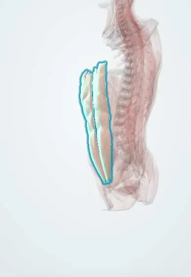
Rectus abdominis !
The rectus abdominis is the "six-pack" muscle down the front of the abdomen, from the ribs to the pubis; it bends the trunk forward.
Official name: Rectus abdominis muscle
Opens in the Durgical app, no sign-up needed.
What it is
The 'six-pack' muscle running down the front of the abdomen from the ribs to the pubis.
What it does
Bends the trunk forward (as in a sit-up) and tilts the pelvis back. The ALIF incision goes beside or through its sheath.
Key facts
- Origin
- Pubic symphysis and pubic crest
- Insertion
- Xiphoid process and costal cartilages 5–7
- Action
- Flexes trunk (lumbar spine); compresses abdomen; tilts pelvis posteriorly
- Nerve
- Thoracoabdominal nerves (T7–T11) and subcostal nerve (T12)
- Blood supply
- Superior and inferior epigastric arteries
Relations
- In front
- Anterior rectus sheath, fixed at the tendinous intersections; the small pyramidalis lies in front of its lower end in many people
- Behind
- Above the arcuate line: posterior rectus sheath (internal oblique and transversus abdominis aponeuroses); below it: transversalis fascia and peritoneum only
- Medial
- Linea alba, the midline seam between the two muscles
- Lateral
- Linea semilunaris, where the flat muscles' aponeuroses meet the sheath
- Contents
- Within the sheath: superior and inferior epigastric vessels on its deep surface and the ends of the T7–T12 nerves, which enter from the side
How to find it
Lying on the back and lifting the head and shoulders, the two muscles stand out beside the midline, with the tendinous intersections as horizontal grooves. Its lateral edge, the linea semilunaris, meets the right costal margin near the tip of the ninth costal cartilage, the surface marking of the gallbladder.
Examination
- Beevor sign: on a head lift from lying flat, an upward shift of the navel shows weakness of the lower rectus abdominis (lesion near T10, or some myopathies)
- Carnett sign: abdominal pain that stays the same or worsens when the wall is tensed by a head lift points to a source in the wall itself rather than inside the abdomen
On imaging
Ultrasound and CT show the paired muscles in their sheath beside the linea alba. Rectus diastasis is a widened gap between them without a fascial defect, best seen on ultrasound while the patient lifts the head. A rectus sheath haematoma is a lens-shaped mass inside the sheath; below the arcuate line it can cross the midline and reach the bladder. CT shows active bleeding.
Common problems
- Delayed or weak activation is seen in some people with chronic low back pain.
How it is diagnosed
Exam; ultrasound can show its activation.
Treatment
- Core stability exercise programmes.
Clinical notes
- Rectus diastasis: the linea alba stretches and the two muscles drift apart, common after pregnancy; a midline ridge appears on sitting up, but the wall is stretched, not breached as in a hernia
- Rectus sheath haematoma: a torn epigastric vessel, more often below the arcuate line and in people on anticoagulants, gives a painful wall mass that stays on one side of the midline
- Rectus abdominis flaps (TRAM, VRAM) on the epigastric vessels are used to rebuild the breast and to fill perineal and pelvic defects
- Incisional hernias after a midline laparotomy come through the linea alba between the two muscles
Anatomical variants
- The number of tendinous intersections varies: three is usual, but two or four occur, and one below the navel is often incomplete
Development
Develops from thoracic myotomes that grow forward around the trunk wall, shifting from a lateral to a front position; the tendinous intersections are thought to be leftovers of that segmentation, and its nerve supply stays segmental (T7–T12).
Related conditions
- Incisional (ventral) hernia
- Gastroschisis and omphalocele
- Surgical site infection
- Wound dehiscence and evisceration
- Abdominal compartment syndrome
Test yourself
Think of the answer, then tap the question to see it.
What are the attachments of the rectus abdominis?
From the pubic symphysis and pubic crest to the xiphoid process and costal cartilages 5–7.
How does the rectus sheath change below the arcuate line?
All three aponeuroses pass in front of the rectus abdominis; only transversalis fascia lies behind it.
What is the Beevor sign?
On a head lift, the navel moves up because the lower rectus abdominis is weak, classically from a lesion near T10.
Which arteries meet inside the rectus abdominis?
The superior and inferior epigastric arteries, linking the subclavian and external iliac systems.
How does rectus diastasis differ from a hernia?
In diastasis the linea alba stretches and the two rectus abdominis muscles drift apart; the wall is stretched, not breached.
Educational information, not medical advice. For any symptom, see a health professional.
Its names
- Official Latin (TA2)
- musculus rectus abdominis TA2 2357
- Official Spanish (DeCS)
- Recto del abdomen DeCS D017568
- English (TA2)
- Rectus abdominis muscle
Related
Sources: Terminologia Anatomica (TA2); DeCS; Anatomy, Anterolateral Abdominal Wall Muscles; Anatomy of the Human Body (20th ed.): Development of the Muscles (Lea & Febiger (Gray's Anatomy, 1918), 1918) · adapted, Public domain; 11.4 Axial Muscles of the Abdominal Wall, and Thorax (OpenStax (Rice University), 2022) · adapted, CC BY 4.0.
Spotted a mistake?We are always improving for the community. Tell us what is wrong and our team will review it.Report a mistake
Representative model, for educational purposes only.