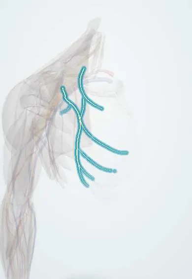
Pectoral nerves !
The pectoral nerves, medial and lateral, reach the chest muscles from the brachial plexus; they move the pectoralis major and minor.
Official name: Medial and lateral pectoral nerves
Opens in the Durgical app, no sign-up needed.
What it is
Nerves from the cords to the chest muscles.
What it does
Power pectoralis major and minor.
Key facts
- Roots
- Lateral C5–C7 (lateral cord); medial C8–T1 (medial cord)
- Moves
- Pectoralis major (both); pectoralis minor (medial)
- Feeling from
- None
Relations
- Course
- The lateral pectoral nerve leaves the lateral cord, crosses in front of the axillary artery and pierces the clavipectoral fascia to the deep surface of pectoralis major; the medial pectoral nerve leaves the medial cord, runs between the axillary artery and vein and pierces pectoralis minor, supplying it and the lower pectoralis major
- In front
- Pectoralis major, which both nerves enter on its deep surface
- Behind
- The axillary artery, in front of which a loop joins the two nerves (ansa pectoralis)
- Deep
- Pectoralis minor and the clavipectoral fascia
How to find it
Not palpable. On lifting pectoralis major they are seen on its deep surface: the lateral pectoral nerve with the thoracoacromial vessels above pectoralis minor, the medial pectoral nerve coming through or around pectoralis minor. On ultrasound the lateral pectoral nerve lies beside the pectoral branch of the thoracoacromial artery, between the two pectoral muscles.
Examination
- Clavicular head: resisted horizontal adduction with the arm raised above shoulder level; sternocostal head: palms pressed together in front of the waist; a head that does not tense under the fingers suggests loss of its nerve supply
On imaging
Not seen on radiographs or routine CT. On ultrasound they are found beside the pectoral branch of the thoracoacromial artery in the plane between pectoralis major and minor, the target of the PECS block used in breast surgery. After nerve injury, MRI shows oedema and later fatty atrophy of the pectoral muscles.
Clinical notes
- In mastectomy and level II–III axillary clearance, retracting or dividing pectoralis minor puts the medial pectoral nerve at risk; its loss wastes the lower sternocostal part of pectoralis major
- Interpectoral (Rotter) lymph nodes lie between the two pectoral muscles close to the lateral pectoral nerve and vessels
- Pectoral nerve blocks between the pectoral muscles and over serratus anterior give analgesia for breast and anterior chest wall surgery
- The lateral or medial pectoral nerve can be used as a donor in nerve transfers to the musculocutaneous or axillary nerve
Anatomical variants
- The medial pectoral nerve usually comes from the medial cord but may pass around the lower border of pectoralis minor instead of through it
- The size of the loop between the two nerves varies, and either nerve may be split into several branches
Development
Pectoralis major and minor are limb-derived muscles that spread over the front of the chest, not body-wall muscles, so the plexus supplies them. The nerves are named for their cords, not their position: the medial pectoral nerve ends up lateral to the lateral one.
Memory aid
'Lateral is medial, medial is lateral': the names follow the cords, not where the nerves lie on the chest wall.
Test yourself
Think of the answer, then tap the question to see it.
Why does the lateral pectoral nerve lie medial to the medial pectoral nerve on the chest wall?
The pectoral nerves are named for the cord they come from (lateral and medial cords), not their position.
What does each pectoral nerve pierce on its way to pectoralis major?
The lateral pectoral nerve pierces the clavipectoral fascia with the thoracoacromial artery; the medial pectoral nerve pierces pectoralis minor.
Which pectoral nerve is at risk when pectoralis minor is retracted in axillary clearance, and with what result?
The medial pectoral nerve; its loss wastes the lower sternocostal part of pectoralis major.
Educational information, not medical advice. For any symptom, see a health professional.
Its names
- Official Latin (TA2)
- nervus pectoralis medialis TA2 6444
- Official Spanish (DeCS)
- Nervios pectorales medial y lateral
- English (TA2)
- Medial and lateral pectoral nerves
Related
Sources: Terminologia Anatomica (TA2); Anatomy, Thorax, Medial Pectoral Nerves; Anatomy, Shoulder and Upper Limb, Pectoral Muscles; Anatomy of the Human Body (20th ed.): Development of the Muscles (Lea & Febiger (Gray's Anatomy, 1918), 1918) · adapted, Public domain.
Spotted a mistake?We are always improving for the community. Tell us what is wrong and our team will review it.Report a mistake
Representative model, for educational purposes only.