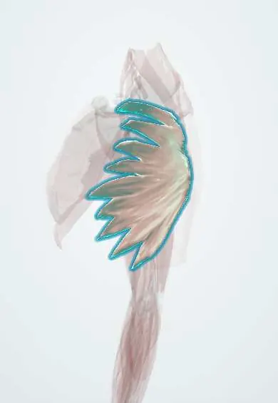
Serratus anterior !
The serratus anterior wraps the side of the ribcage to the shoulder blade; it holds the blade flat on the ribs and swings it forward to reach or push.
Opens in the Durgical app, no sign-up needed.
What it is
A saw-toothed muscle on the side of the ribs, running under the shoulder blade to its inner edge.
What it does
Holds the shoulder blade flat on the ribs and swings it forward and up when you reach or push.
Key facts
- Origin
- External surfaces of lateral parts of ribs 1–8 (varies: 1–9)
- Insertion
- Anterior surface of medial border of scapula, mostly at inferior angle
- Action
- Protracts scapula and holds it against chest wall; rotates it upward for arm elevation above 90°
- Nerve
- Long thoracic nerve (C5–C7); injury gives winged scapula
- Blood supply
- Lateral thoracic artery; thoracodorsal artery
Relations
- Deep
- Ribs 1–8 or 9 and the external intercostal muscles of the lateral chest wall
- Superficial
- Long thoracic nerve and lateral thoracic artery on its outer surface; pectoralis major and pectoralis minor in front, latissimus dorsi over its lower back part
- Behind
- Subscapularis, separated by the loose scapulothoracic gliding plane
- Lateral
- The axilla, whose medial wall it forms, with the axillary lymph nodes of the chest wall group along it
How to find it
With the arm raised forward and the hand pushing against a wall, the saw-tooth slips stand out on the side of the chest below the armpit, between the pectoralis major and the latissimus dorsi; clearest in lean, muscular people.
Examination
- Wall push-up: the medial border and inferior angle of the scapula lift off the chest wall; this points to serratus anterior weakness, usually a long thoracic nerve palsy
- Resisted forward flexion or a forward punch: medial winging that appears on flexion, rather than on abduction, places the problem in the serratus anterior rather than the trapezius
On imaging
On axial CT or MRI it lies on the outer surface of the ribs along the side of the chest, deep to the scapula. Long thoracic nerve palsy is diagnosed clinically (medial winging) and by electromyography; MRI shows atrophy and fatty change later. Ultrasound shows it over the ribs in the mid-axillary line and guides the serratus anterior plane block.
Common problems
- Winged scapula: the inner edge of the shoulder blade lifts off the ribs when the long thoracic nerve is injured.
- Weakness makes overhead reaching and pushing hard and can strain the shoulder.
How it is diagnosed
Wall push-up test shows the winging. EMG checks the long thoracic nerve.
Treatment
- Physical therapy; most nerve stretch injuries recover over months.
- Muscle or tendon transfer surgery if the nerve does not recover.
Clinical notes
- Long thoracic nerve palsy also follows heavy shoulder straps, repetitive overhead sport and neuralgic amyotrophy; most traction palsies recover slowly
- Without it the scapula cannot rotate up and stay on the chest wall, so elevation of the arm stops short and scapulothoracic rhythm breaks down
- Its lower slips, on the serratus branch of the thoracodorsal artery, are used as a free flap alone or with a latissimus dorsi flap; sparing the upper slips avoids winging
Anatomical variants
- The number of rib slips varies; the lowest can reach the 9th or even the 10th rib
Development
It forms from premuscle tissue in the lower neck and migrates a long way down the side of the chest; its nerve, the long thoracic nerve from roots C5–C7, is drawn out with it, which explains its long exposed course.
Memory aid
Long thoracic nerve: 'C5, 6, 7 raise the wings to heaven'.
Related conditions
Test yourself
Think of the answer, then tap the question to see it.
What nerve supplies the serratus anterior, and where is it exposed?
The long thoracic nerve (C5–C7), on its outer surface along the mid-axillary line; at risk in axillary node dissection, mastectomy and chest drain insertion.
What sign does serratus anterior weakness give, and how is it brought out?
Medial winging: on a wall push-up the medial border and inferior angle of the scapula lift off the chest wall.
Why does arm elevation stop short when the serratus anterior is paralysed?
The scapula can no longer rotate upward and stay on the chest wall, so scapulothoracic rhythm breaks down.
Which wall of the axilla does the serratus anterior form?
The medial wall.
Educational information, not medical advice. For any symptom, see a health professional.
Its names
- Official Latin (TA2)
- musculus serratus anterior TA2 2307
- Official Spanish (DeCS)
- Músculo serrato anterior
- English (TA2)
- Serratus anterior
Related
Sources: Terminologia Anatomica (TA2); Anatomy, Thorax, Serratus Anterior Muscles; Anatomy of the Human Body (20th ed.): Development of the Muscles (Lea & Febiger (Gray's Anatomy, 1918), 1918) · adapted, Public domain.
Spotted a mistake?We are always improving for the community. Tell us what is wrong and our team will review it.Report a mistake
Representative model, for educational purposes only.