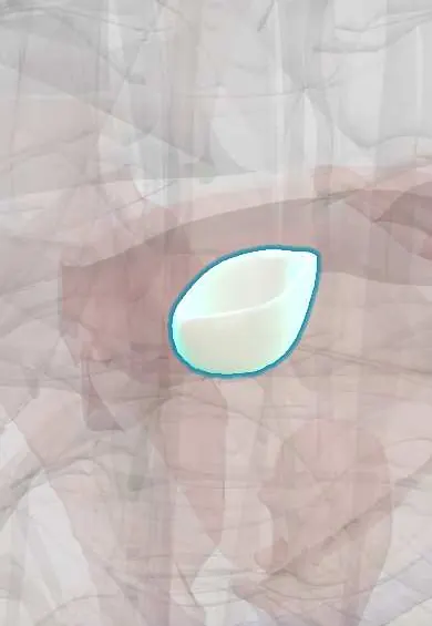
Aortic non-coronary cusp !
The non-coronary cusp is the one flap of the aortic valve with no coronary artery above it; it sits close to the heart's main wiring.
Official name: Non-coronary leaflet of aortic valve
Opens in the Durgical app, no sign-up needed.
What it is
The aortic cusp with no coronary artery above it, at the back.
What it does
One of the three cusps that seal the aortic valve.
Key facts
- Where
- Right posterior cusp of aortic valve; no coronary artery arises from its sinus; faces interatrial septum
- Attaches
- Crescent-shaped attachment to aortic root; nodule and lunules at free edge; sinus of Valsalva behind it
- Function
- Closes in diastole, preventing backflow into left ventricle; valve heard best at right 2nd intercostal space
Relations
- Behind
- Interatrial septum, with the right atrium and the left atrium on either side
- In front
- Aortic right coronary cusp and aortic left coronary cusp, meeting it at the commissures
- Below
- Mitral anterior leaflet (shared aortomitral curtain) and, at the commissure with the right cusp, the membranous septum
How to find it
In the opened aortic root it is the posterior cusp, lying over the interatrial septum between the two atria; it is the only sinus with no coronary opening. On transesophageal echo it is the cusp next to the septum in the short-axis view.
On imaging
Parasternal short-axis echo and transesophageal echo show it next to the interatrial septum, between the two atria; endocarditis abscesses around it appear as echo-free or thickened areas in the root.
Common problems
- Aortic stenosis: calcium stiffens the cusps with age; causes chest pain, fainting and breathlessness. Severe disease needs replacement.
- Bicuspid valve: born with two cusps instead of three (about 1-2% of people), wears out earlier.
- Aortic regurgitation (leak).
How it is diagnosed
Murmur on exam; echocardiogram measures the valve area and the pressure drop; CT for valve sizing before TAVR.
Treatment
- Valve replacement: open surgery or a catheter valve (TAVR) placed through an artery in the groin.
Clinical notes
- After transcatheter aortic valve implantation, new left bundle branch block or complete heart block comes from pressure on conduction tissue below the right–non-coronary commissure
- Endocarditis abscess here can burrow into the right atrium or the interatrial septum and cause AV block on the ECG
Development
It comes from the posterior cushion of the outflow tract, the one swelling that is not split when the outflow septum divides the two lateral cushions between the aorta and the pulmonary trunk.
Related conditions
Test yourself
Think of the answer, then tap the question to see it.
Why is the aortic non-coronary cusp so named, and what does it face?
No coronary artery arises from its sinus; it faces the interatrial septum, between the right and left atria.
Why can transcatheter aortic valve implantation cause left bundle branch block or complete heart block?
Pressure on conduction tissue below the right–non-coronary commissure, which lies over the membranous septum and the bundle of His.
Where does a sinus of Valsalva aneurysm of the non-coronary sinus typically rupture?
Into the right atrium; the non-coronary sinus is the second commonest site after the right sinus.
Educational information, not medical advice. For any symptom, see a health professional.
Its names
- Official Latin (TA2)
- valvula noncoronaria TA2 3997
- Official Spanish (DeCS)
- Valva semilunar no coronaria DeCS D000082902
- English (TA2)
- Non-coronary leaflet of aortic valve
- Also called
- posterior semilunar leaflet of aortic valve · nonadjacent leaflet of aortic valve · velum semilunare dextrum · posterior semilunar cusp · noncoronary cusp
Related
Sources: Terminologia Anatomica (TA2); DeCS; Anatomy, Thorax, Aortic Valve; Anatomy of the Human Body (20th ed.): Development of the Vascular System (Lea & Febiger (Gray's Anatomy, 1918), 1918) · adapted, Public domain.
Spotted a mistake?We are always improving for the community. Tell us what is wrong and our team will review it.Report a mistake
Representative model, for educational purposes only.