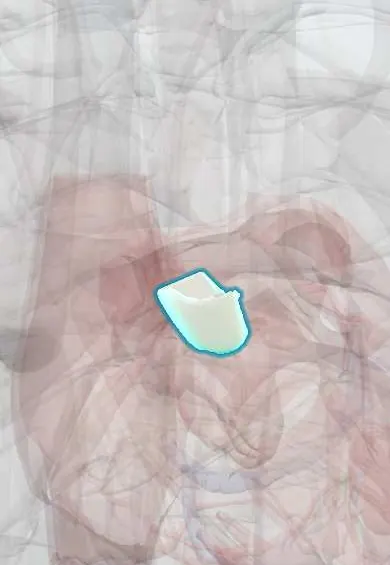
Aortic right coronary cusp !
The right coronary cusp is one of the three flaps of the aortic valve, the front one; the right coronary artery starts just above it.
Official name: Right coronary leaflet
Opens in the Durgical app, no sign-up needed.
What it is
The aortic cusp with the opening of the right coronary artery above it.
What it does
One of the three cusps that seal the aortic valve.
Key facts
- Where
- Anterior cusp of aortic valve; its sinus gives rise to right coronary artery; valve behind left sternum at 3rd intercostal space
- Attaches
- Crescent-shaped attachment to aortic root; nodule and lunules at free edge; sinus of Valsalva behind it
- Function
- Closes in diastole, preventing backflow into left ventricle; valve heard best at right 2nd intercostal space
Relations
- In front
- Right ventricular outflow tract (infundibulum), with the pulmonary trunk in front and to the left
- Behind
- Aortic left coronary cusp and aortic non-coronary cusp, meeting it at the commissures
- Below
- Membranous septum below its commissure with the non-coronary cusp, with the bundle of His just underneath
How to find it
Aortic area: right 2nd intercostal space beside the sternum. Aortic regurgitation is often louder at the left 3rd to 4th intercostal space, with the person sitting forward and breath held after breathing out.
On imaging
Parasternal short axis at the base: the closed cusps form an inverted Y, with this cusp in front. CT before transcatheter valve implantation measures how high the coronary ostia sit above the annulus.
Common problems
- Aortic stenosis: calcium stiffens the cusps with age; causes chest pain, fainting and breathlessness. Severe disease needs replacement.
- Bicuspid valve: born with two cusps instead of three (about 1-2% of people), wears out earlier.
- Aortic regurgitation (leak).
How it is diagnosed
Murmur on exam; echocardiogram measures the valve area and the pressure drop; CT for valve sizing before TAVR.
Treatment
- Valve replacement: open surgery or a catheter valve (TAVR) placed through an artery in the groin.
Clinical notes
- Sinus of Valsalva aneurysm most often affects the right coronary sinus and can rupture into the right ventricle or right atrium
- An anomalous coronary artery from the wrong sinus running between the aorta and pulmonary trunk is a cause of sudden death in young athletes
- A perimembranous or subarterial ventricular septal defect can suck this cusp into the defect (prolapse), causing aortic regurgitation
Anatomical variants
- Bicuspid aortic valve in about 1% of people, usually with unequal cusps; fusion of the right and left coronary cusps is the commonest pattern
Development
The aortic and pulmonary cusps grow from swellings on the outflow tract cushions as the truncus arteriosus divides; abnormal fusion of two swellings gives a bicuspid valve.
Memory aid
Auscultation areas from base to apex: 'All Physicians Take Money' (Aortic right 2nd space, Pulmonic left 2nd space, Tricuspid left lower sternal border, Mitral apex).
Related conditions
- Syncope (vasovagal, orthostatic hypotension, cardiac)
- Aortic Valve Stenosis
- Infective endocarditis
- Aortic regurgitation
Test yourself
Think of the answer, then tap the question to see it.
Which coronary artery arises from the sinus of the aortic right coronary cusp?
The right coronary artery, which runs forward and right to the atrioventricular groove.
Where is the murmur of aortic stenosis heard, and how does it sound?
A crescendo–decrescendo systolic murmur at the right 2nd intercostal space, spreading to the carotids.
Which sinus of Valsalva is most often affected by aneurysm, and where does it rupture?
The right coronary sinus; it can rupture into the right ventricle or the right atrium.
In a bicuspid aortic valve, which cusps are most often fused?
The right and left coronary cusps, the commonest pattern.
Educational information, not medical advice. For any symptom, see a health professional.
Its names
- Official Latin (TA2)
- valvula coronaria dextra TA2 3995
- Official Spanish (DeCS)
- Valva semilunar coronaria derecha DeCS D000082902
- English (TA2)
- Right coronary leaflet
- Also called
- right semilunar leaflet of aortic valve · velum semilunare ventrale · right semilunar cusp
Related
Sources: Terminologia Anatomica (TA2); DeCS; Anatomy, Thorax, Aortic Valve.
Spotted a mistake?We are always improving for the community. Tell us what is wrong and our team will review it.Report a mistake
Representative model, for educational purposes only.