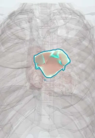
Left atrium !
The left atrium is the upper left chamber of the heart, at its back; it receives oxygen-rich blood from the lungs and fills the left ventricle.
Opens in the Durgical app, no sign-up needed.
What it is
The upper left chamber of the heart, which receives oxygen-rich blood from the lungs through four pulmonary veins.
What it does
Holds blood from the lungs and tops up the left ventricle through the mitral valve. Its small pouch (the left atrial appendage) is where most clots form in atrial fibrillation.
Key facts
- Where
- Forms most of base (posterior surface) of heart; receives four pulmonary veins; esophagus lies directly behind
- Blood supply
- Circumflex artery (atrial branches); some right coronary branches
- Nerve
- Cardiac plexus: sympathetic (T1–T4) speeds and strengthens; vagus nerves slow
- Function
- Receives oxygenated blood from lungs and fills left ventricle through mitral valve
Relations
- In front
- Roots of the ascending aorta and the pulmonary trunk, separated from it by the transverse pericardial sinus
- Behind
- Oblique pericardial sinus, then the esophagus and the descending thoracic aorta
- Above
- Tracheal bifurcation and the left main bronchus above its roof; the right pulmonary artery passes over it
- Below
- Mitral valve opening into the left ventricle; the coronary sinus runs in the posterior atrioventricular groove
- Lateral
- Left atrial appendage curving forward along the left side of the pulmonary trunk, close to the circumflex artery
- Medial
- Interatrial septum, with the right atrium beyond it
How to find it
It cannot be felt or heard directly. Mitral murmurs that reflect its pressure are heard at the apex (mitral area: left 5th intercostal space, midclavicular line), best with the person turned onto the left side and with the bell for low-pitched diastolic sounds.
On imaging
Chest X-ray: an enlarged left atrium gives a double contour behind the right heart border, a bulge on the left border below the pulmonary trunk, a wider carinal angle and a backward-pushed barium-filled esophagus. Echo: parasternal long axis for size; transesophageal echo for the appendage and clots.
Common problems
- Atrial fibrillation: the chamber quivers instead of contracting, and a clot can form and travel to the brain (stroke).
- Enlarges with a leaking or narrowed mitral valve or high blood pressure.
How it is diagnosed
ECG for the rhythm; echocardiogram for size and clots (transesophageal echo looks at the appendage).
Treatment
- Blood thinners to prevent stroke, rate or rhythm medicines, catheter ablation, or closing the appendage with a device.
Clinical notes
- Rheumatic mitral stenosis raises left atrial pressure: atrial fibrillation, lung congestion and, rarely, hoarseness when the big atrium presses on the left recurrent laryngeal nerve (Ortner syndrome)
- Transesophageal echo before cardioversion looks for thrombus in the appendage; the appendage can be closed or excluded in selected patients
- Catheter ablation for atrial fibrillation isolates the pulmonary veins where they enter this chamber; the esophagus right behind can suffer thermal injury (atrioesophageal fistula)
- A fold of atrial wall separates the appendage opening from the left upper pulmonary vein (left lateral ridge); on transesophageal echo it can look like a thrombus
- Bachmann bundle, a muscle band crossing the atrial roofs from near the superior vena cava to the left appendage, is the main fast route between the atria; its block widens and notches the P wave (interatrial block)
Anatomical variants
- Pulmonary veins: four separate openings are usual, but the two left veins often join in a common trunk and an extra right middle vein is fairly frequent; these patterns are mapped on CT before ablation
- The appendage varies in shape (chicken wing, windsock, cactus, cauliflower); some studies link the shapes other than chicken wing with more strokes
Development
Its smooth wall comes from the primitive pulmonary vein absorbed into the atrium; only the appendage keeps the rough, pectinate wall of the primitive atrium.
Related conditions
- Mitral stenosis
- Supraventricular tachycardia (AVNRT, AVRT)
- Restrictive cardiomyopathy (amyloid, sarcoid, haemochromatosis)
- Cardiac tumours (atrial myxoma)
- Atrial fibrillation
- Hypertensive heart disease
- Rheumatic heart disease
- Atrial septal defect
Test yourself
Think of the answer, then tap the question to see it.
Why does transesophageal echo show the left atrium and its appendage best?
The left atrium is the most posterior chamber of the heart and lies directly in front of the esophagus.
Where do most clots form in atrial fibrillation, and what is the main risk?
In the left atrial appendage; a clot that breaks off can travel to the brain and cause a stroke.
What is Ortner syndrome?
Hoarseness when a very large left atrium, as in rheumatic mitral stenosis, presses on the left recurrent laryngeal nerve.
Which complication of catheter ablation for atrial fibrillation comes from the relations of the left atrium?
Atrioesophageal fistula: the esophagus lies right behind the left atrium where the pulmonary veins are isolated, so it can suffer thermal injury.
Which chest X-ray signs suggest an enlarged left atrium?
A double contour behind the right heart border, a bulge on the left border below the pulmonary trunk and a wider carinal angle.
Educational information, not medical advice. For any symptom, see a health professional.
Its names
- Official Latin (TA2)
- atrium sinistrum; auricula sinistra cordis TA2 4054
- Official Spanish (DeCS)
- Atrio izquierdo DeCS D006325
- English (TA2)
- Left atrium
- Also called
- left atrial appendage
Related
Sources: Terminologia Anatomica (TA2); DeCS; Anatomy, Thorax, Heart; Anatomy, Thorax, Heart and Pericardial Cavity; Tratado de anatomía humana (9.ª ed.), tomo II, Libro IV Angiología, sección 1, capítulo 1: Corazón, artículos I–VI.
Spotted a mistake?We are always improving for the community. Tell us what is wrong and our team will review it.Report a mistake
Representative model, for educational purposes only.