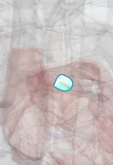
Aortic left coronary cusp !
The left coronary cusp is one of the three flaps of the aortic valve; the left main coronary artery starts just above it.
Official name: Left coronary leaflet
Opens in the Durgical app, no sign-up needed.
What it is
The aortic cusp with the opening of the left coronary artery above it.
What it does
One of the three cusps that seal the aortic valve.
Key facts
- Where
- Left posterior cusp of aortic valve; its sinus gives rise to left main coronary artery
- Attaches
- Crescent-shaped attachment to aortic root; nodule and lunules at free edge; sinus of Valsalva behind it
- Function
- Closes in diastole, preventing backflow into left ventricle; valve heard best at right 2nd intercostal space
Relations
- In front
- Aortic right coronary cusp, at the commissure facing the pulmonary valve; the pulmonary trunk lies in front and to the left
- Behind
- Transverse pericardial sinus, with the left atrium behind it
- Below
- Aortomitral curtain and the anterior mitral leaflet
- Lateral
- Left atrial appendage and the left main coronary artery heading left
How to find it
Aortic area at the right 2nd intercostal space; the stenotic murmur spreads to both carotids, where a weak, slow-rising pulse can be felt in severe disease.
On imaging
CT and coronary angiography show the left main ostium in this sinus; on parasternal short-axis echo the cusp lies to the left and back, beside the left atrial appendage.
Common problems
- Aortic stenosis: calcium stiffens the cusps with age; causes chest pain, fainting and breathlessness. Severe disease needs replacement.
- Bicuspid valve: born with two cusps instead of three (about 1-2% of people), wears out earlier.
- Aortic regurgitation (leak).
How it is diagnosed
Murmur on exam; echocardiogram measures the valve area and the pressure drop; CT for valve sizing before TAVR.
Treatment
- Valve replacement: open surgery or a catheter valve (TAVR) placed through an artery in the groin.
Clinical notes
- Coronary obstruction during transcatheter aortic valve implantation most often involves the left main, when the ostium is low and the sinus shallow
- Endocarditis on this cusp can seed the anterior mitral leaflet through the shared fibrous curtain
Anatomical variants
- In bicuspid valves, right–left coronary cusp fusion is commonest, then right–non-coronary; left–non-coronary fusion is rare
Development
It comes from half of a lateral cushion of the outflow tract, split by the septum that divides the truncus into aorta and pulmonary trunk. The coronary arteries grow in later and open into the left and right sinuses.
Related conditions
Test yourself
Think of the answer, then tap the question to see it.
Which artery arises from the sinus of the aortic left coronary cusp, and how does it divide?
The left main coronary artery; it runs behind the pulmonary trunk and divides into the LAD and the circumflex artery.
Which pulse and heart sound findings suggest severe aortic stenosis?
A weak, slow-rising carotid pulse (pulsus parvus et tardus) and a soft or single second heart sound.
Which coronary is most often obstructed during transcatheter aortic valve implantation, and when?
The left main, when its ostium in the left coronary sinus is low and the sinus is shallow.
Educational information, not medical advice. For any symptom, see a health professional.
Its names
- Official Latin (TA2)
- valvula coronaria sinistra TA2 3996
- Official Spanish (DeCS)
- Valva semilunar coronaria izquierda DeCS D000082902
- English (TA2)
- Left coronary leaflet
- Also called
- left semilunar leaflet of aortic valve · velum semilunare sinistrum · left semilunar cusp
Sources: Terminologia Anatomica (TA2); DeCS; Anatomy, Thorax, Aortic Valve; Anatomy of the Human Body (20th ed.): Development of the Vascular System (Lea & Febiger (Gray's Anatomy, 1918), 1918) · adapted, Public domain.
Spotted a mistake?We are always improving for the community. Tell us what is wrong and our team will review it.Report a mistake
Representative model, for educational purposes only.