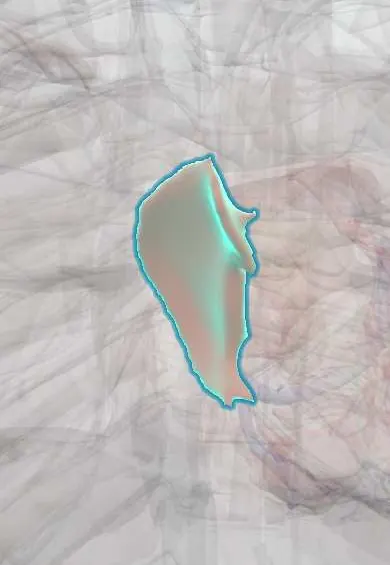
Interatrial septum !
The interatrial septum is the thin wall between the two upper chambers of the heart; its oval dip marks an opening that closed after birth.
Opens in the Durgical app, no sign-up needed.
What it is
The thin wall between the two atria, with a shallow dip (the fossa ovalis) where a fetal opening closed after birth.
What it does
Separates the atria. In about 1 in 4 adults the fetal flap is not fully sealed (patent foramen ovale).
Key facts
- Where
- Between atria; fossa ovalis with limbus on right atrial side
- Function
- Separates atria; fossa ovalis is remnant of fetal foramen ovale (right-to-left shunt); probe-patent in about 25% of adults
Relations
- In front
- Aortic root at the aortic non-coronary cusp, bulging against its front part
- Above
- Opening of the superior vena cava, where a sinus venosus defect lies
- Below
- Inferior vena cava and coronary sinus openings; below lies the atrioventricular septum with the triangle of Koch and the Atrioventricular (AV) node
- Behind
- Interatrial groove on the back of the heart, behind which the right pulmonary veins enter the left atrium; surgeons open this groove to reach the left atrium and mitral valve
How to find it
From the opened right atrium it is the oval dip on the medial wall, above the inferior vena cava opening, with the limbus felt as a ridge. In the catheter laboratory the fossa ovalis is the thinnest point and the site of transseptal puncture into the left atrium.
On imaging
Echocardiography: subcostal views in children and transesophageal echo in adults show it best; colour Doppler and an agitated saline (bubble) study show a shunt. A large atrial septal defect dilates the right atrium and right ventricle and enlarges the pulmonary arteries on X-ray.
Common problems
- Patent foramen ovale, usually harmless; sometimes closed with a device after certain strokes.
- Atrial septal defect (a true hole).
How it is diagnosed
Echocardiogram; ECG for rhythm problems.
Treatment
- Treatment of the underlying cause; catheter ablation for atrial flutter.
Clinical notes
- Transseptal puncture through the fossa ovalis gives catheter access to the left atrium for ablation, mitral procedures and appendage closure
- Selected secundum defects and some patent foramen ovale after certain strokes are closed with a catheter device; primum and sinus venosus defects lack rims and are repaired surgically
- A large unrepaired atrial septal defect leads to right heart dilation, atrial arrhythmias and, late, pulmonary hypertension with shunt reversal (Eisenmenger syndrome)
Anatomical variants
- Atrial septal aneurysm: a floppy fossa ovalis that bulges from side to side, often with a patent foramen ovale
- The fossa ovalis varies in shape (oval, round or elliptical) and size
Development
The septum primum grows down and its upper part breaks open (ostium secundum); the septum secundum grows to its right, leaving the foramen ovale. At birth, higher left atrial pressure presses the two together.
Related conditions
Test yourself
Think of the answer, then tap the question to see it.
What forms the floor and the rim (limbus) of the fossa ovalis?
The floor is the old septum primum; the limbus is the edge of the septum secundum.
What is a patent foramen ovale, and how can it cause paradoxical embolism?
A flap that never sealed (probe-patent in about 25% of adults); it opens when right atrial pressure rises, as with cough or straining.
Which atrial septal defect is commonest, and what heart sound does it give?
The secundum defect, at the fossa ovalis; it gives a wide, fixed split second heart sound.
Where is transseptal puncture made to reach the left atrium?
Through the fossa ovalis of the interatrial septum, its thinnest point.
Educational information, not medical advice. For any symptom, see a health professional.
Its names
- Official Latin (TA2)
- septum interatriale; fossa ovalis atrii dextri TA2 3964
- Official Spanish (DeCS)
- Tabique interatrial DeCS D054087
- English (TA2)
- Interatrial septum
- Also called
- oval fossa of right atrium · atrial septum · septum atriorum
Related
Sources: Terminologia Anatomica (TA2); DeCS; Anatomy, Thorax, Heart Fossa Ovalis; Anatomy, Thorax, Heart Foramen Ovale; Tratado de anatomía humana (9.ª ed.), tomo II, Libro IV Angiología, sección 1, capítulo 1: Corazón, artículos I–VI.
Spotted a mistake?We are always improving for the community. Tell us what is wrong and our team will review it.Report a mistake
Representative model, for educational purposes only.