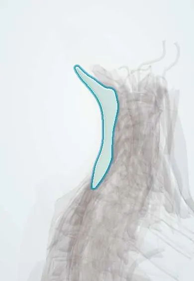
Nuchal ligament !
The nuchal ligament is a thin midline sheet at the back of the neck, from the skull to C7; it helps hold the head up and anchors neck muscles.
Opens in the Durgical app, no sign-up needed.
What it is
A thin midline sheet at the back of the neck, from the skull to C7: the upward continuation of the supraspinous ligament.
What it does
Helps hold the head up and anchors neck muscles such as the trapezius and splenius.
Key facts
- Attaches
- External occipital protuberance and crest to posterior tubercle of atlas and spinous processes C2–C7
- Function
- Midline septum for muscle attachment (trapezius, splenius capitis, rhomboid minor, serratus posterior superior); helps support the head
- Tight in
- Neck and head flexion
Relations
- Superficial
- Skin and fat of the back of the neck, and the trapezius origin on each side
- Deep
- Posterior tubercle of the atlas, spinous processes of C2–C7 and the interspinous ligaments; at the top, the posterior atlanto-occipital membrane
- Lateral
- Splenius capitis and semispinalis capitis; the greater occipital nerve comes out through semispinalis capitis and trapezius a little lateral to its upper end
How to find it
With the chin tucked to the chest it tightens and is felt as a midline ridge from the external occipital protuberance down to the prominent C7 spinous process.
On imaging
A lateral cervical radiograph sometimes shows a small calcification (nuchal ossicle) behind the spinous processes, an incidental finding. MRI can show high signal after a flexion or whiplash injury.
Clinical notes
- The midline posterior cervical approach follows this plane, limiting bleeding and muscle damage
- Clay-shoveler fracture: avulsion of a lower cervical or upper thoracic spinous process (C7 most often) by the pull of the muscles and ligaments attached to it
Anatomical variants
- Calcified or ossified nodules inside the ligament, usually in the lower neck, are a frequent incidental finding on lateral neck X-rays; they should not be mistaken for a fractured spinous process
Related conditions
Test yourself
Think of the answer, then tap the question to see it.
Where does the nuchal ligament attach?
From the external occipital protuberance and crest to the posterior tubercle of the atlas and the spinous processes of C2–C7; it continues the supraspinous ligament upward.
Which muscles attach to the nuchal ligament?
The trapezius, splenius capitis, rhomboid minor and serratus posterior superior; it serves as a midline septum for them.
Why does the midline posterior cervical approach follow the nuchal ligament?
It divides the back of the neck into right and left muscle halves, a relatively bloodless plane that limits bleeding and muscle damage.
What is a clay-shoveler fracture, near the lower end of the nuchal ligament?
Avulsion of a lower cervical or upper thoracic spinous process, most often C7, by the pull of the muscles and ligaments attached to it.
Educational information, not medical advice. For any symptom, see a health professional.
Its names
- Official Latin (TA2)
- ligamentum nuchae TA2 1678
- Official Spanish (DeCS)
- Ligamento nucal
- English (TA2)
- Nuchal ligament
- Also called
- septum nuchae
Related
Sources: Terminologia Anatomica (TA2); Anatomy, Head and Neck: Cervical Vertebrae; Anatomy, Head and Neck, Suboccipital Muscles.
Spotted a mistake?We are always improving for the community. Tell us what is wrong and our team will review it.Report a mistake
Representative model, for educational purposes only.