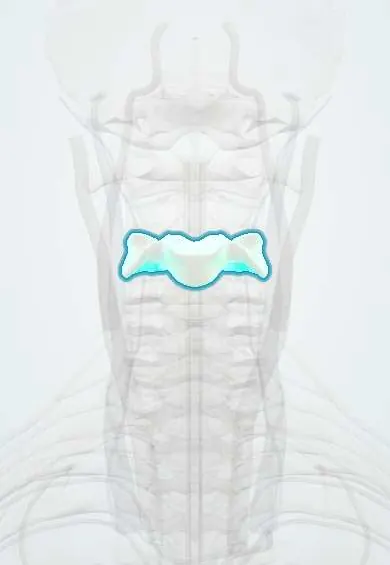
C3 vertebra !
C3 is the third neck (cervical) vertebra; with the others it holds up the head, protects the spinal cord and lets the neck move.
Official name: C3
Opens in the Durgical app, no sign-up needed.
What it is
The C3 neck vertebra: a small body in front, a ring behind for the spinal cord, and holes on each side for the vertebral artery.
What it does
Stacks with the other neck vertebrae to hold up the head, protect the spinal cord and let the neck bend and turn.
Key facts
- Joints with
- C2 above and C4 below (intervertebral discs, facet joints and uncovertebral joints)
- Landmarks
- Small wide body with uncinate processes; transverse foramina (vertebral artery); short bifid spinous process; level of hyoid bone
Relations
- In front
- Longus colli and longus capitis, the prevertebral fascia and the retropharyngeal space, then the pharynx
- Lateral
- Vertebral artery and vertebral vein in the transverse foramina; the C4 root runs out in the gutter of the transverse process behind the artery; scalenes and levator scapulae attach to the tubercles
- Behind
- Short bifid spinous process under the nuchal ligament, with semispinalis, multifidus and splenius layered over the laminae
- Contents
- Spinal cord at roughly the C4 segment: cervical cord segments sit about one level above the vertebra with the same number
How to find it
With the neck slightly flexed, the C2 spinous process is found below the occiput; C3 lies just below it, but its spine is short and set deep in the cervical curve, so it is often hard to feel. In front, the level matches the hyoid bone, felt above the thyroid cartilage.
On imaging
Lateral X-ray: the soft tissue in front of C2–C4 is normally thin (commonly quoted under about 7 mm in adults); a thicker shadow suggests a hematoma from a hidden fracture or a retropharyngeal abscess. Oblique views or CT show foramina narrowed by uncovertebral spurs.
Common problems
- Wear and bone spurs (cervical spondylosis), most often at C5-C6 and C6-C7.
- Spurs or a disc can narrow the canal (pressing the cord) or a side opening (pinching a nerve root).
- Fractures from falls, car accidents or diving.
How it is diagnosed
X-rays show alignment and spurs. MRI shows discs, nerves and the spinal cord. CT shows bone detail.
Treatment
- Most neck pain settles with time, activity changes, physical therapy and pain medicine.
- Surgery if a nerve or the cord is pressed and symptoms are severe or getting worse, for example ACDF.
Clinical notes
- Retropharyngeal abscess in young children spreads in the space in front of these vertebrae and widens the prevertebral shadow
- Injuries of the subaxial spine (C3–C7) are scored by fracture shape, the state of the disc and ligaments, and neurology (SLIC system)
- Injury of the C3–C5 segments can paralyse the diaphragm on one or both sides; a raised hemidiaphragm on the chest film is the clue
- The superior cervical sympathetic ganglion lies on the longus capitis in front of the upper cervical transverse processes (about C2–C3); stretching or cutting it during a high anterior exposure can leave Horner syndrome on that side
Development
Each typical cervical vertebra ossifies from three primary centres (body and each half of the arch). Failure of segmentation gives block vertebrae; C2–C3 is a frequent site in Klippel–Feil syndrome (short neck, low hairline, limited movement).
Memory aid
"C3, 4, 5 keeps the diaphragm alive": the phrenic nerve roots.
Test yourself
Think of the answer, then tap the question to see it.
Which joints do the uncinate processes of the C3 vertebra form, and why do they matter clinically?
The uncovertebral joints; their spurs narrow the intervertebral foramen behind them.
Which spinal nerve exits above the C3 vertebra?
The C3 nerve: in the neck each root leaves above the vertebra with the same number, so the C4 nerve exits below C3.
Why can a spinal cord injury at or above the C3 vertebra leave a patient dependent on a ventilator?
It threatens the phrenic nerve output from C3–C5 to the diaphragm («C3, 4, 5 keeps the diaphragm alive»).
Which anterior neck landmark lies at the level of the C3 vertebra?
The hyoid bone, felt above the thyroid cartilage.
Educational information, not medical advice. For any symptom, see a health professional.
Its names
- Official Latin (TA2)
- vertebra cervicalis III TA2 1032
- Official Spanish (DeCS)
- Vértebra cervical C3 DeCS D002574
- English (TA2)
- C3
Related
- C2–C3 disc
- C3–C4 disc
- Vertebral artery
- Anterior longitudinal ligament
- Endplates
- Neck facet joints
- Interspinales
- Interspinous ligaments
Sources: Terminologia Anatomica (TA2); DeCS; Anatomy, Head and Neck: Cervical Vertebrae; Anatomy, Back, Cervical Vertebrae.
Spotted a mistake?We are always improving for the community. Tell us what is wrong and our team will review it.Report a mistake
Representative model, for educational purposes only.