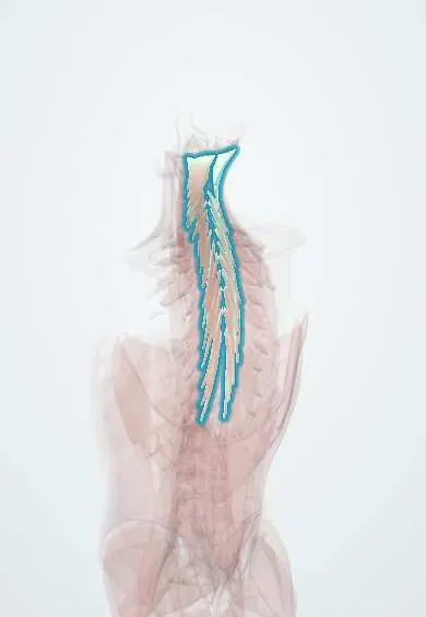
Semispinalis !
The semispinalis is a deep back muscle under the erector spinae, thickest in the neck; it straightens the spine and holds the head up.
Official name: Semispinalis muscles
Opens in the Durgical app, no sign-up needed.
What it is
A deep muscle under the erector spinae, crossing four to six vertebrae; in the neck it is a thick muscle up to the skull.
What it does
Straightens the thoracic spine and neck and holds the head up; it turns the trunk slightly to the other side.
Key facts
- Origin
- Transverse processes of C4–T12 (capitis: C4–C7 articular and T1–T6 transverse processes)
- Insertion
- Spinous processes 4–6 segments above (thoracis, cervicis up to C2); capitis: occipital bone between superior and inferior nuchal lines
- Action
- Extends head, cervical and thoracic spine; unilateral: rotates to the opposite side
- Nerve
- Dorsal rami of cervical and thoracic spinal nerves (capitis: greater occipital nerve, C2)
- Blood supply
- Occipital, deep cervical and posterior intercostal arteries
Relations
- Superficial
- Splenius capitis and cervicis, then trapezius
- Deep
- The suboccipital triangle (vertebral artery, suboccipital nerve) beneath the capitis part; multifidus and rotatores beneath the thoracic and cervical parts
- Lateral
- Longissimus capitis and cervicis
- Medial
- Nuchal ligament and the spinous processes, separating left and right
How to find it
Lying face down and lifting the head against light resistance, the two rounded columns stand out on each side of the nuchal furrow just below the occiput, felt through the thin upper trapezius.
On imaging
On axial MRI or CT of the neck, semispinalis capitis is the large paired muscle beside the nuchal ligament, deep to trapezius and splenius, with semispinalis cervicis beneath it. Ultrasound at C2 shows the greater occipital nerve between semispinalis capitis and obliquus capitis inferior, a common block site. After whiplash, MRI can show strain or haematoma as oedema.
Common problems
- Muscle strain and spasm: one of the most common causes of sudden low back pain.
- Weakness and fatigue add to chronic back pain.
How it is diagnosed
History and exam; imaging is not needed for a simple strain.
Treatment
- Staying active, heat, pain medicine and an exercise programme. Most strains settle in days to weeks.
Clinical notes
- A greater occipital nerve block is given where the nerve surfaces near the occiput after piercing semispinalis capitis and trapezius, about a third of the way from the external occipital protuberance to the mastoid
- Detaching semispinalis cervicis from C2 in posterior cervical surgery is linked to loss of the neck's curve and axial neck pain, so surgeons try to preserve or reattach it
Development
An epaxial muscle formed from several fused dorsal myotomes; this is why its supply, like that of the skin over the back of the scalp, comes from dorsal rami, including the greater occipital nerve (C2) that passes through it.
Related conditions
Test yourself
Think of the answer, then tap the question to see it.
Which is the largest muscle at the back of the neck?
The semispinalis capitis, the rounded column on each side of the nuchal furrow under the trapezius and splenius.
Which nerve pierces the semispinalis capitis, and what can its entrapment cause?
The greater occipital nerve (dorsal ramus of C2); entrapment there is a possible cause of occipital neuralgia.
Which way does the semispinalis rotate the spine when it acts on one side?
To the opposite side; bilaterally it extends the head and the cervical and thoracic spine.
Why do surgeons try to preserve the semispinalis cervicis attachment to C2?
Detaching it in posterior cervical surgery is linked to loss of the neck's curve and axial neck pain.
Educational information, not medical advice. For any symptom, see a health professional.
Its names
- Official Latin (TA2)
- musculi semispinales TA2 2280
- Official Spanish (DeCS)
- Músculo semiespinoso DeCS D064170
- English (TA2)
- Semispinalis muscles
- Also called
- semispinalis muscle
Related
Sources: Terminologia Anatomica (TA2); DeCS; Anatomy, Back; Anatomy of the Human Body (20th ed.): Development of the Muscles (Lea & Febiger (Gray's Anatomy, 1918), 1918) · adapted, Public domain.
Spotted a mistake?We are always improving for the community. Tell us what is wrong and our team will review it.Report a mistake
Representative model, for educational purposes only.