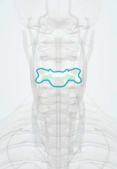
C4 vertebra !
C4 is the fourth neck (cervical) vertebra; with the others it holds up the head, protects the spinal cord and lets the neck move.
Official name: C4
Opens in the Durgical app, no sign-up needed.
What it is
The C4 neck vertebra: a small body in front, a ring behind for the spinal cord, and holes on each side for the vertebral artery.
What it does
Stacks with the other neck vertebrae to hold up the head, protect the spinal cord and let the neck bend and turn.
Key facts
- Joints with
- C3 above and C5 below (intervertebral discs, facet joints and uncovertebral joints)
- Landmarks
- Small wide body with uncinate processes; transverse foramina (vertebral artery); short bifid spinous process; level of upper thyroid cartilage and carotid bifurcation (C3–C4)
Relations
- In front
- Prevertebral fascia and longus colli, then the laryngopharynx and larynx; anterolaterally the carotid sheath with the carotid arteries, the internal jugular vein and the vagus nerve
- Lateral
- Vertebral artery in the transverse foramen; the C5 root leaves through the C4–C5 foramen and passes between the anterior and middle scalenes to the upper trunk of the brachial plexus
- Behind
- Bifid spine under the nuchal ligament; levator scapulae arises from the posterior tubercles of the upper four transverse processes
- Contents
- Spinal cord at the top of the cervical enlargement (segments C4–T1, which supply the upper limbs)
How to find it
In front it lies at about the upper border of the thyroid cartilage. From behind its short spine is deep and hard to pick out; counting up from the prominent C7 is more reliable than counting down from C2.
On imaging
Lateral X-ray: count down from C2 and check the four smooth lines (front and back of the bodies, spinolaminar line, spinous tips) for steps. In central cord syndrome MRI may show cord swelling or bleeding at a narrowed spondylotic level with no fracture.
Common problems
- Wear and bone spurs (cervical spondylosis), most often at C5-C6 and C6-C7.
- Spurs or a disc can narrow the canal (pressing the cord) or a side opening (pinching a nerve root).
- Fractures from falls, car accidents or diving.
How it is diagnosed
X-rays show alignment and spurs. MRI shows discs, nerves and the spinal cord. CT shows bone detail.
Treatment
- Most neck pain settles with time, activity changes, physical therapy and pain medicine.
- Surgery if a nerve or the cord is pressed and symptoms are severe or getting worse, for example ACDF.
Clinical notes
- Anterior cervical approaches at upper levels put the superior laryngeal and hypoglossal nerves at risk; lower levels put the recurrent laryngeal nerve at risk
- The carotid sinus sits at the bifurcation in front of this level: pressure there slows the heart (the basis of carotid sinus massage), and a hypersensitive sinus can cause fainting
- Facet dislocations of the lower neck: a forward slip of about a quarter of the body depth suggests one jumped facet, about half suggests both, with a high risk of cord injury
Development
The neck is part of the single forward-bent curve of the fetus; the cervical lordosis is a secondary curve that forms after birth, once the infant starts holding the head up. Its apex usually lies around C4–C5, and its loss on a lateral film is a common finding after injury or with degeneration.
Related conditions
Test yourself
Think of the answer, then tap the question to see it.
Which nerve root leaves through the C4–C5 foramen below the C4 vertebra, and what does its compression weaken?
The C5 nerve root: weak deltoid and biceps, with a reduced biceps reflex.
What is central cord syndrome at the level of the C4 vertebra in an older adult?
A cord injury without fracture from hyperextension of a spondylotic neck: the arms are weaker than the legs, and the hands are worst.
Why is irritation of the diaphragm felt at the top of the shoulder, at the C4 level?
The C4 dermatome caps the top of the shoulder, and the phrenic nerve carries diaphragm pain to that segment (shoulder-tip pain).
Educational information, not medical advice. For any symptom, see a health professional.
Its names
- Official Latin (TA2)
- vertebra cervicalis IV TA2 1032
- Official Spanish (DeCS)
- Vértebra cervical C4 DeCS D002574
- English (TA2)
- C4
Related
- C3–C4 disc
- C4–C5 disc
- Vertebral artery
- Anterior longitudinal ligament
- Dura (dural sac)
- Endplates
- Neck facet joints
- Interspinales
Sources: Terminologia Anatomica (TA2); DeCS; Anatomy, Head and Neck: Cervical Vertebrae; Anatomy, Back, Cervical Vertebrae; 7.3 The Vertebral Column (OpenStax (Rice University), 2022) · adapted, CC BY 4.0.
Spotted a mistake?We are always improving for the community. Tell us what is wrong and our team will review it.Report a mistake
Representative model, for educational purposes only.