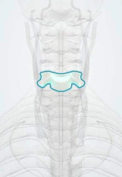
C5 vertebra !
C5 is the fifth neck vertebra, a typical one: a small body in front and a ring behind that protects the spinal cord.
Official name: C5
Opens in the Durgical app, no sign-up needed.
What it is
A typical neck vertebra: a small body in front, a ring behind for the spinal cord, and holes on each side for the vertebral artery.
What it does
Stacks with the other neck vertebrae to hold up the head, protect the spinal cord and let the neck bend and turn.
Key facts
- Joints with
- C4 above and C6 below (intervertebral discs, facet joints and uncovertebral joints)
- Landmarks
- Small wide body with uncinate processes; transverse foramina (vertebral artery); short bifid spinous process; C5–C6 is the most common level of cervical spondylosis
Relations
- In front
- Longus colli and the prevertebral fascia, then the laryngopharynx and the larynx; the carotid sheath lies anterolaterally
- Lateral
- Vertebral artery in the transverse foramen; the C6 nerve root leaves through the C5–C6 foramen behind the artery, between the uncovertebral joint in front and the facet joint behind
- Behind
- Neck facet joints and laminae under semispinalis and multifidus; the ligamentum flavum lines the back of the canal
- Contents
- Spinal cord at about the C6 segment, within the cervical enlargement; narrowing here is the classic site of spondylotic myelopathy
How to find it
In front it lies behind the thyroid cartilage (Adam's apple). From behind, the short spine is felt only with the neck flexed, counting up two from C7.
Examination
- Spurling test: extension and side-bending toward the painful side with gentle downward pressure on the head; arm pain along a dermatome suggests foraminal root compression
- Hoffmann sign: flicking the nail of the middle finger makes the thumb and index finger flex; a sign of upper motor neuron (cord) compression
On imaging
Lateral view: disc height loss and front and back spurs at C5–C6; canal size can be compared with body width (Torg–Pavlov ratio, below about 0.8 suggests a narrow canal). MRI shows cord compression and T2 signal change in myelopathy; oblique views or CT show foraminal spurs.
Common problems
- Wear and bone spurs (cervical spondylosis), most often at C5-C6 and C6-C7.
- Spurs or a disc can narrow the canal (pressing the cord) or a side opening (pinching a nerve root).
- Fractures from falls, car accidents or diving.
How it is diagnosed
X-rays show alignment and spurs. MRI shows discs, nerves and the spinal cord. CT shows bone detail.
Treatment
- Most neck pain settles with time, activity changes, physical therapy and pain medicine.
- Surgery if a nerve or the cord is pressed and symptoms are severe or getting worse, for example ACDF.
Clinical notes
- Anterior cervical discectomy and fusion reaches the spine between the carotid sheath laterally and the trachea and esophagus medially; the recurrent laryngeal nerve is the nerve at risk
- Cervical myelopathy shows as clumsy hands and unsteady gait with brisk reflexes, most often from multilevel spondylosis centred on C5–C6
- Facet dislocations favour C5–C6 and C6–C7, often after flexion with rotation in road crashes or falls
Anatomical variants
- Developmentally narrow cervical canal: on a lateral X-ray the canal is small in relation to the body (Torg–Pavlov ratio under about 0.8); it leaves little room for the cord, raising the risk of transient arm and leg weakness after a tackle and of myelopathy with age
Development
After the primary centres have joined, small secondary centres appear around puberty at the tips of the spinous and transverse processes and as rings on the upper and lower rims of the body; they fuse in early adulthood, and an unfused tip can be mistaken for a small fracture.
Related conditions
Test yourself
Think of the answer, then tap the question to see it.
Which nerve root does a C5–C6 disc or spur below the C5 vertebra compress, and what are the signs?
The C6 nerve root: weak elbow flexion and wrist extension, reduced biceps and brachioradialis reflexes, and a numb thumb.
What is a flexion teardrop fracture, most often seen at the C5 vertebra?
A triangular fragment breaks off the front of the body while the rest is pushed back into the canal; it is highly unstable, often with an anterior cord syndrome.
In neck and arm pain at the level of the C5 vertebra, what does a positive Spurling test suggest?
Foraminal nerve root compression: extension and side-bending toward the painful side with gentle downward pressure reproduce arm pain along a dermatome.
Which nerve is at risk in anterior cervical discectomy and fusion at the C5 vertebra?
The recurrent laryngeal nerve; the approach passes between the carotid sheath laterally and the trachea and esophagus medially.
Educational information, not medical advice. For any symptom, see a health professional.
Its names
- Official Latin (TA2)
- vertebra cervicalis V TA2 1032
- Official Spanish (DeCS)
- Vértebra cervical C5 DeCS D002574
- English (TA2)
- C5
Related
- C4–C5 disc
- C5–C6 disc
- Vertebral artery
- Anterior longitudinal ligament
- Dura (dural sac)
- Endplates
- Neck facet joints
- Interspinales
Sources: Terminologia Anatomica (TA2); DeCS; Anatomy, Head and Neck: Cervical Vertebrae; Anatomy, Back, Cervical Vertebrae.
Spotted a mistake?We are always improving for the community. Tell us what is wrong and our team will review it.Report a mistake
Representative model, for educational purposes only.