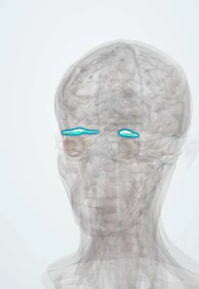
Levator palpebrae !
The levator palpebrae is a thin muscle in the eye socket that ends in the upper eyelid; it lifts the lid, which droops when the muscle or its nerve fails.
Official name: Levator palpebrae superioris
Opens in the Durgical app, no sign-up needed.
What it is
A thin muscle above the superior rectus, ending in the upper eyelid.
What it does
Lifts the upper eyelid; weak in a third-nerve palsy (droopy lid).
Key facts
- Origin
- Lesser wing of the sphenoid above the optic canal
- Insertion
- Skin and tarsal plate of the upper eyelid
- Action
- Elevates the upper eyelid
- Nerve
- Oculomotor nerve (III), superior division; superior tarsal (Müller) part: sympathetic
- Blood supply
- Ophthalmic artery (muscular branches)
Relations
- Course
- From the lesser wing of the sphenoid it runs forward under the orbital roof, above the superior rectus, to Whitnall ligament; the aponeurosis then descends in the upper eyelid, its horns anchored to the medial and lateral orbital walls
- Above
- Orbital roof; the frontal nerve and supraorbital vessels run forward on its upper surface
- Below
- Superior rectus, which shares its fascial sheath and the superior division of the oculomotor nerve, so the lid rises with upgaze
- In front
- Orbital septum, with the preaponeurotic fat pad between septum and aponeurosis: the surgeon's landmark in ptosis repair
- Deep
- Superior tarsal (Müller) muscle and the conjunctiva of the upper fornix
- Lateral
- Lateral horn of the aponeurosis, which divides the lacrimal gland into orbital and palpebral lobes
How to find it
Its action is judged by the movement of the upper lid. The examiner holds the brow still with a thumb (to cancel frontalis) and watches the lid margin from full downgaze to full upgaze. The lid crease, where aponeurotic fibres reach the skin, lies a few millimetres above the margin.
Examination
- Levator function: lid excursion from downgaze to upgaze with the brow fixed, normally about 15 mm; poor function (about 4 mm or less) suggests a congenital (myogenic) or neurogenic cause
- Margin reflex distance 1 (MRD1): from the corneal light reflex to the upper lid margin, normally about 4–5 mm; it grades how severe the ptosis is
- Fatigue and ice-pack tests: ptosis that worsens with sustained upgaze and improves after the lid is cooled suggests myasthenia gravis
- Pupils in every ptosis: a large pupil on the side of the ptosis points to a compressive oculomotor (III) palsy, a small one to Horner syndrome
On imaging
On coronal CT or MRI it lies just above the superior rectus under the orbital roof and is hard to separate from it; sagittal MRI follows it forward into the aponeurosis of the upper lid. Ptosis itself is judged clinically; imaging seeks the cause: MRI with MR or CT angiography in a third nerve palsy with a dilated pupil (aneurysm), or a search along the sympathetic chain in Horner syndrome.
Clinical notes
- A pupil-involving oculomotor (III) palsy with ptosis and an eye turned down and out can signal a posterior communicating artery aneurysm, an emergency; pupil-sparing palsies are often microvascular (diabetes, hypertension)
- Congenital ptosis that covers the pupil can cause amblyopia and is corrected early, often with a frontalis sling when levator function is poor
- Aponeurotic ptosis is also seen after eye surgery and with long-term contact lens wear; repair reattaches or advances the aponeurosis on the tarsus
- Aberrant regeneration after an oculomotor (III) palsy: fibres meant for other muscles reach the levator, so the lid rises when the eye looks down or inward
- Thyroid eye disease causes lid retraction and lid lag on downgaze, from overaction of the superior tarsal muscle and scarring of the levator
Development
It splits off from the superior rectus primordium during development, which is why congenital ptosis may come with weak elevation of the same eye.
Related conditions
Test yourself
Think of the answer, then tap the question to see it.
Which muscles lift the upper eyelid, and what is the nerve supply of each?
The levator palpebrae superioris (oculomotor nerve III) does most; the superior tarsal (Müller) muscle adds about 1–2 mm under sympathetic control.
How does ptosis differ between an oculomotor (III) palsy and Horner syndrome?
Oculomotor palsy: complete ptosis (levator); Horner syndrome: mild ptosis with a small pupil, as only the Müller muscle is lost.
How is levator function measured, and what is normal?
Lid excursion from downgaze to upgaze with the brow held still; normally about 15 mm.
What are the features of aponeurotic ptosis in older adults?
The levator aponeurosis thins or detaches from the tarsus; levator function stays good and the lid crease rises.
What can a pupil-involving oculomotor (III) palsy with ptosis signal?
A posterior communicating artery aneurysm, an emergency; pupil-sparing palsies are often microvascular.
Educational information, not medical advice. For any symptom, see a health professional.
Its names
- Official Latin (TA2)
- levator palpebrae superioris TA2 2052
- Official Spanish (DeCS)
- Músculo elevador del párpado superior DeCS D009801
- English (TA2)
- Levator palpebrae superioris
- Also called
- levator palpebrae superioris muscle · lamina superficialis levatoris palpebrae superioris
Related
Sources: Terminologia Anatomica (TA2); DeCS; Anatomy, Head and Neck: Levator Palpebrae Superioris Muscle.
Spotted a mistake?We are always improving for the community. Tell us what is wrong and our team will review it.Report a mistake
Representative model, for educational purposes only.