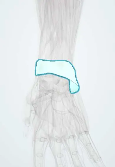
Flexor retinaculum !
The flexor retinaculum is the tough band across the front of the wrist that roofs the carpal tunnel, where the median nerve and finger flexors pass.
Opens in the Durgical app, no sign-up needed.
What it is
A thick band across the palm side of the wrist bones that forms the roof of the carpal tunnel.
What it does
Keeps the finger flexor tendons close to the wrist when it bends.
Key facts
- Attaches
- Medially pisiform and hook of the hamate; laterally scaphoid tubercle and trapezium crest
- Function
- Roof of the carpal tunnel; pulley for the flexor tendons
- Tight in
- Wrist flexion (tendons bowstring against it)
Relations
- Contents
- Carpal tunnel: median nerve, FDS tendons, FDP tendons and FPL tendon, wrapped in the ulnar and radial bursae
- Superficial
- Palmaris longus tendon, the palmar cutaneous branch of the median nerve and, distally, the palmar aponeurosis; the ulnar nerve and ulnar artery in Guyon canal at its ulnar edge
- Deep
- Floor of the tunnel: carpal bones covered by the palmar wrist ligaments and midcarpal ligaments
- Lateral
- FCR tendon in its own compartment against the trapezium; the recurrent motor branch of the median nerve curls up to the thenar muscles near the distal edge
- Below
- Beyond the distal edge, the superficial palmar arch and the common digital nerves, both at risk if a release goes too far
How to find it
The retinaculum lies just distal to the distal wrist crease at the base of the palm, between the thenar and hypothenar eminences. Its ulnar pillars (pisiform, hook of the hamate) and radial pillars (scaphoid tubercle, trapezium ridge) can be felt as firm points at its four corners.
Examination
- Phalen test: the wrists are held fully flexed for about a minute; tingling in the thumb, index and middle fingers suggests carpal tunnel syndrome
- Tinel sign at the wrist: tapping over the tunnel sends tingling into the median nerve fingers
- Durkan carpal compression test: the examiner presses with the thumbs directly over the tunnel for about 30 seconds; reproduction of the symptoms is positive
- Thenar wasting and weak palmar abduction of the thumb (APB) point to longstanding compression of the recurrent motor branch
On imaging
On ultrasound the retinaculum is a thin echogenic band spanning the pisiform–scaphoid level to the hamate–trapezium level; the median nerve's cross-sectional area is measured at the tunnel inlet. MRI can show palmar bowing of the retinaculum and, after release, its divided edges.
Common problems
- Carpal tunnel syndrome: the tunnel under it gets crowded and squeezes the median nerve.
- Night tingling in the thumb, index and middle fingers; later numbness and thumb weakness.
How it is diagnosed
Symptoms, tapping (Tinel) and wrist-bending (Phalen) tests. Nerve conduction studies and EMG measure how badly the nerve is squeezed; ultrasound can show a swollen nerve.
Treatment
- Night wrist splint, activity changes and sometimes a steroid injection.
- Carpal tunnel release: cutting this ligament to give the nerve room, open or endoscopic.
Clinical notes
- Carpal tunnel release divides the retinaculum along its ulnar side, in line with the radial border of the ring finger, away from the recurrent motor branch and the palmar cutaneous branch
- Distal limit of the release: the fat pad and superficial palmar arch just beyond the distal edge, near Kaplan's cardinal line
- Pillar pain: tenderness over the thenar and hypothenar bases for weeks or months after release
- Hook of hamate fractures (golf, bat and racquet sports) involve its ulnar attachment and can irritate the ulnar nerve or fray the little-finger flexor tendons
- Acute carpal tunnel syndrome after distal radius fractures or perilunate dislocations can need urgent decompression
Anatomical variants
- The recurrent motor branch usually leaves the median nerve beyond the distal edge, but may arise within the tunnel or pierce the retinaculum, a risk during release
- A bifid median nerve, sometimes with a persistent median artery between its two parts
Related conditions
Test yourself
Think of the answer, then tap the question to see it.
What are the attachments of the flexor retinaculum?
Medially the pisiform and hook of the hamate; laterally the scaphoid tubercle and the trapezium crest.
What passes through the carpal tunnel under the flexor retinaculum?
The median nerve and nine tendons: four FDS, four FDP and the FPL; the FCR runs in its own split, outside the tunnel.
Why is thenar skin sensation spared in carpal tunnel syndrome?
The palmar cutaneous branch of the median nerve passes superficial to the flexor retinaculum.
What is the Phalen test?
The wrists are held fully flexed for about a minute; tingling in the thumb, index and middle fingers suggests carpal tunnel syndrome.
Along which side is the flexor retinaculum divided in carpal tunnel release, and why?
Its ulnar side, in line with the radial border of the ring finger, away from the recurrent motor branch and the palmar cutaneous branch.
Educational information, not medical advice. For any symptom, see a health professional.
Its names
- Official Latin (TA2)
- retinaculum flexorium carpi TA2 2550
- Official Spanish (DeCS)
- Retináculo de los músculos flexores
- English (TA2)
- Flexor retinaculum
- Also called
- transverse carpal ligament
Related
- Median nerve
- FDP tendons
- Superficial palmar arch
- Recurrent motor branch
- Palmar cutaneous branch
- Palmar aponeurosis
- Pisiform ligaments
- Carpal tunnel
Sources: Terminologia Anatomica (TA2); Anatomy, Shoulder and Upper Limb, Wrist Flexor Retinaculum.
Spotted a mistake?We are always improving for the community. Tell us what is wrong and our team will review it.Report a mistake
Representative model, for educational purposes only.