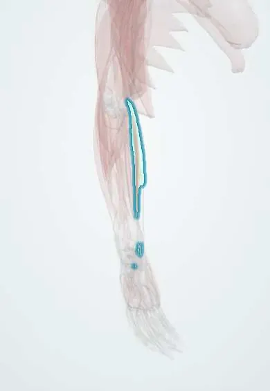
Flexor carpi radialis !
The flexor carpi radialis runs down the front of the forearm to the thumb side of the hand; it bends the wrist and tilts it toward the thumb.
Opens in the Durgical app, no sign-up needed.
What it is
A forearm muscle from the inner elbow to the base of the index metacarpal.
What it does
Bends the wrist and tilts it toward the thumb.
Key facts
- Origin
- Medial epicondyle (common flexor tendon)
- Insertion
- Bases of the 2nd and 3rd metacarpals
- Action
- Flexes and abducts (radially deviates) the wrist
- Nerve
- Median nerve (C6–C7)
- Blood supply
- Radial and ulnar arteries
Relations
- Course
- From the common flexor tendon it runs obliquely across the forearm, becomes tendinous halfway down and passes through a separate canal in the trapezium groove, lateral to the carpal tunnel, to the bases of the 2nd and 3rd metacarpals
- Lateral
- Pronator teres above; at the wrist the radial artery and further out the brachioradialis tendon
- Medial
- Palmaris longus (when present) and, at the wrist, the median nerve
- Deep
- Flexor digitorum superficialis (FDS) above; in the distal forearm flexor pollicis longus (FPL) and pronator quadratus
How to find it
With the wrist flexed against resistance and tilted slightly toward the thumb, its tendon stands out at the wrist crease as the cord lateral to palmaris longus; the radial pulse is felt just on its thumb side.
Examination
- Resisted wrist flexion with radial deviation: pain along the tendon near the scaphoid tubercle and trapezium suggests FCR tendinitis; weakness with a normal flexor carpi ulnaris suggests a high median nerve lesion
On imaging
Ultrasound first. In a transverse view at the wrist the tendon lies radial to the median nerve, then enters its own tunnel in the groove of the trapezium. Tenosynovitis shows as fluid and a thick sheath; a tear as a gap. MRI if ultrasound is unclear. Radiographs do not show it, but show scaphotrapeziotrapezoid arthritis beside its tunnel, a common irritant.
Clinical notes
- Volar approach to the distal radius: the FCR sheath is opened, the tendon retracted, FPL swept aside and pronator quadratus lifted; the radial artery is protected laterally and the median nerve medially
- FCR tendinitis in its tight trapezium tunnel, often with arthritis of the scaphotrapeziotrapezial joint
- Volar wrist ganglia often sit between the FCR tendon and the radial artery, so excision puts the artery at risk
- Donor for transfer to the finger extensors (extensor digitorum) in permanent radial nerve palsy
Related conditions
Test yourself
Think of the answer, then tap the question to see it.
What lies on either side of the flexor carpi radialis tendon at the wrist?
The radial artery pulse just lateral, and the median nerve just medial and deep.
Does the flexor carpi radialis tendon run in the carpal tunnel?
No; it runs in its own fibro-osseous tunnel in the groove of the trapezium, to the bases of the 2nd and 3rd metacarpals.
Which surgical approach to the distal radius uses the flexor carpi radialis sheath?
The volar (FCR, modified Henry) approach: the sheath is opened, FPL swept aside and pronator quadratus lifted.
Educational information, not medical advice. For any symptom, see a health professional.
Its names
- Official Latin (TA2)
- flexor radialis carpi TA2 2481
- Official Spanish (DeCS)
- Músculo flexor radial del carpo
- English (TA2)
- Flexor carpi radialis
Related
Sources: Terminologia Anatomica (TA2); Anatomy, Shoulder and Upper Limb, Forearm Muscles; Anatomy, Shoulder and Upper Limb, Forearm Compartments.
Spotted a mistake?We are always improving for the community. Tell us what is wrong and our team will review it.Report a mistake
Representative model, for educational purposes only.