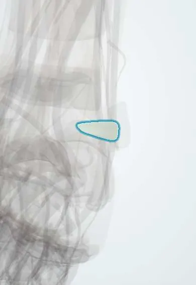
Joints and ligaments · Lower limb
PTFL !
The posterior talofibular ligament is the strongest ligament of the outer ankle, at the back; it is rarely torn.
Official name: Posterior talofibular ligament
Opens in the Durgical app, no sign-up needed.
What it is
A thick ligament at the back of the outer ankle, from the fibula to the talus.
What it does
Keeps the talus from slipping backward. It is the strongest of the three outer ankle ligaments.
Key facts
- Attaches
- Malleolar fossa of fibula to lateral tubercle of posterior process of talus
- Function
- Strongest lateral ligament; resists posterior displacement and external rotation of talus
- Tight in
- Dorsiflexion
Relations
- Above
- Posterior inferior tibiofibular ligament and its deep transverse part, just above it
- Medial
- Flexor hallucis longus tendon in its groove between the lateral and medial tubercles of the talus
- Behind
- Fibularis longus and brevis tendons behind the lateral malleolus; further back, Kager's fat pad and the Achilles tendon
- Below
- CFL, which arises lower down and further forward on the lateral malleolus
How to find it
Not palpable: it lies deep, behind the ankle joint. It is seen through posterior ankle arthroscopy or on MRI.
Examination
- Forced passive plantarflexion test: pain at the back of the ankle suggests posterior impingement (os trigonum or Stieda process) rather than a PTFL injury
On imaging
On axial MRI it normally looks striped (fibres separated by fat); that pattern is not a tear. A lateral X-ray shows an os trigonum or a long lateral tubercle at its attachment.
Common problems
- Rarely torn, except in ankle dislocations.
Clinical notes
- Torn in frank dislocations and in the most severe inversion injuries, together with the ATFL and CFL
- Posterior impingement in ballet dancers and footballers: the lateral tubercle or os trigonum is pinched between the tibia and calcaneus in full plantarflexion
- The posterolateral arthroscopy portal lies near the sural nerve and small saphenous vein
Anatomical variants
- A posterior intermalleolar ligament, lying between this ligament and the inferior transverse ligament, is common and can be trapped in posterior impingement
Development
The lateral tubercle of the talus ossifies from its own centre in late childhood; if it never fuses, it remains a separate os trigonum, which can be mistaken for a fracture.
Test yourself
Think of the answer, then tap the question to see it.
Where does the posterior talofibular ligament (PTFL) attach?
From the malleolar fossa of the fibula to the lateral tubercle of the posterior process of the talus.
In which ankle position is the posterior talofibular ligament tightest?
Dorsiflexion, the opposite of the ATFL.
When is the posterior talofibular ligament usually torn?
Mainly in ankle dislocations; it is the strongest of the three lateral ligaments.
Educational information, not medical advice. For any symptom, see a health professional.
Its names
- Official Latin (TA2)
- ligamentum talofibulare posterius TA2 1920
- Official Spanish (DeCS)
- Ligamento talofibular posterior DeCS D017844
- English (TA2)
- Posterior talofibular ligament
- Also called
- ligamentum fibulotalare posterius · oblique ligament of Walther
Related
Sources: Terminologia Anatomica (TA2); DeCS; Anatomy, Bony Pelvis and Lower Limb: Ankle Joint.
Spotted a mistake?We are always improving for the community. Tell us what is wrong and our team will review it.Report a mistake
Representative model, for educational purposes only.