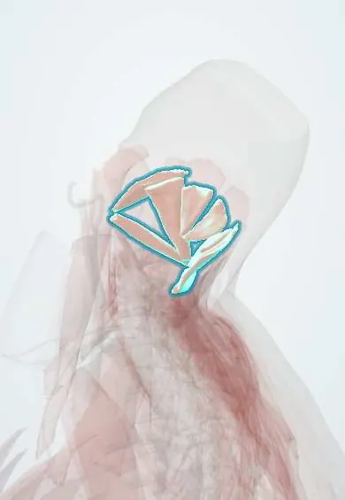
Suboccipital muscles !
The suboccipital muscles are four small muscles deep at the back of the upper neck, between the skull, C1 and C2; they fine-tune head movement.
Official name: Rectus posterior major capitis muscle
Opens in the Durgical app, no sign-up needed.
What it is
Four small muscles between the skull, the atlas and the axis, deep at the back of the upper neck.
What it does
Fine-tune nodding and turning of the head. Tension here is linked to headaches at the base of the skull.
Key facts
- Origin
- Rectus capitis posterior major: C2 spine; minor: C1 posterior tubercle; obliquus capitis inferior: C2 spine; superior: C1 transverse process
- Insertion
- Major: lateral inferior nuchal line; minor: medial inferior nuchal line; inferior oblique: C1 transverse process; superior oblique: occipital bone between the nuchal lines, lateral to rectus capitis posterior major
- Action
- Extend the head; rotate head and atlas to the same side (rectus major, inferior oblique); proprioception
- Nerve
- Suboccipital nerve (dorsal ramus of C1) (some texts: also greater occipital nerve branches, C2, to obliquus capitis inferior)
- Blood supply
- Vertebral and occipital arteries; deep cervical artery
Relations
- Contents
- Suboccipital triangle: the third part of the vertebral artery, the suboccipital venous plexus and the suboccipital nerve
- Superficial
- Semispinalis capitis, then splenius capitis and trapezius; longissimus capitis to the side
- Deep
- Posterior arch of the atlas and the posterior atlanto-occipital membrane, with the dura behind them
- Below
- The greater occipital nerve turning up around obliquus capitis inferior
How to find it
With the person lying on the back and the head resting in the examiner's hands, the fingertips curled under the occiput sink through trapezius and semispinalis into the soft area between the occiput and the C2 spinous process, the first bony point felt below the skull.
On imaging
On axial and sagittal MRI they lie under semispinalis capitis between the occiput, atlas and axis. Ultrasound shows obliquus capitis inferior from the C2 spinous process to the C1 transverse process, with the greater occipital nerve on its surface, the target for an occipital nerve block. CT or MR angiography maps the vertebral artery on the posterior arch of the atlas before surgery.
Clinical notes
- Posterior approaches to C1–C2 and the foramen magnum pass through these muscles; the vertebral artery on top of the posterior arch of the atlas is the structure most at risk
- Compression of the greater occipital nerve where it curves around obliquus capitis inferior and through semispinalis capitis is one proposed cause of occipital neuralgia
- Tender, tight suboccipital muscles are a common finding in tension-type and cervicogenic headache
Anatomical variants
- Rarely an extra rectus capitis muscle or an absent rectus capitis posterior minor (case reports)
Related conditions
Test yourself
Think of the answer, then tap the question to see it.
Which muscles bound the suboccipital triangle?
Rectus capitis posterior major (upper medial), obliquus capitis superior (upper lateral) and obliquus capitis inferior (lower).
What does the suboccipital triangle contain?
The vertebral artery on the posterior arch of the atlas, the suboccipital venous plexus and the suboccipital nerve; its roof is semispinalis capitis.
What nerve supplies the suboccipital muscles?
The suboccipital nerve, the dorsal ramus of C1.
Around which suboccipital muscle does the greater occipital nerve curve?
The lower border of the obliquus capitis inferior, a proposed site of compression in occipital neuralgia.
Educational information, not medical advice. For any symptom, see a health professional.
Its names
- Official Latin (TA2)
- musculus rectus posterior major capitis; musculus rectus posterior minor capitis; musculus obliquus superior capitis; musculus obliquus inferior capitis TA2 2249
- Official Spanish (DeCS)
- Músculos suboccipitales DeCS D009334
- English (TA2)
- Rectus posterior major capitis muscle
- Also called
- rectus capitis posterior major muscle · rectus capitis posterior minor muscle · obliquus capitis superior muscle · obliquus capitis inferior muscle · musculus rectus capitis dorsalis major
Related
Sources: Terminologia Anatomica (TA2); DeCS; Anatomy, Head and Neck, Suboccipital Muscles.
Spotted a mistake?We are always improving for the community. Tell us what is wrong and our team will review it.Report a mistake
Representative model, for educational purposes only.