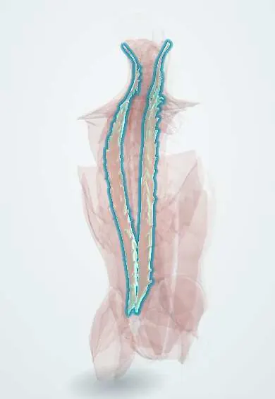
Longissimus !
The longissimus is the middle and longest column of the erector spinae, from the pelvis to the skull base; it straightens the back and neck.
Official name: Longissimus muscles
Opens in the Durgical app, no sign-up needed.
What it is
The middle and longest column of the erector spinae, from the pelvis up to the base of the skull.
What it does
Straightens the back and neck; on one side it bends and turns the head and trunk to that side.
Key facts
- Origin
- Common erector spinae tendon; transverse processes of thoracic and lower cervical vertebrae
- Insertion
- Thoracic and cervical transverse processes; ribs between tubercles and angles; mastoid process (capitis)
- Action
- Bilateral: extends spine and head; unilateral: lateral flexion, turns head to the same side
- Nerve
- Dorsal rami of spinal nerves (lateral branches; cervical, thoracic and lumbar)
- Blood supply
- Dorsal branches of posterior intercostal and lumbar arteries; occipital and deep cervical arteries (capitis)
Relations
- Lateral
- Iliocostalis
- Medial
- Spinalis in the thoracic region; in the lumbar region multifidus, along the Wiltse plane
- Superficial
- Thoracolumbar fascia, latissimus dorsi and the serratus posterior muscles; in the neck, splenius
- Deep
- Transverse processes, levatores costarum and the transversospinal muscles (semispinalis, multifidus)
How to find it
Lying face down with a gentle back extension, it forms most of the paraspinal ridge beside the lumbar spinous processes. On ultrasound for an erector spinae plane block it is the thick muscle seen just above the bright, flat line of a transverse process.
On imaging
On axial MRI or CT it is the large middle column of the erector spinae, between iliocostalis laterally and multifidus medially; the thin fat cleft between it and multifidus is the Wiltse plane. Ultrasound over a transverse process shows it as the thick layer above the bright bony line, where the erector spinae plane block is given. MRI shows strain as oedema, atrophy as fat.
Common problems
- Muscle strain and spasm: one of the most common causes of sudden low back pain.
- Weakness and fatigue add to chronic back pain.
How it is diagnosed
History and exam; imaging is not needed for a simple strain.
Treatment
- Staying active, heat, pain medicine and an exercise programme. Most strains settle in days to weeks.
Clinical notes
- Wiltse approach: going between it and multifidus reaches the facet joints and pedicles without stripping muscle from the midline, used for pedicle screws and far-lateral disc herniations
- Erector spinae plane block: local anaesthetic deep to it spreads up and down several levels to the dorsal and ventral rami, for chest, breast and spine surgery
Development
Epaxial muscle from the dorsal part of the myotomes, so its nerve supply comes from the lateral branches of the dorsal rami. Like the other long columns, it is built from several fused myotomes rather than a single segment.
Memory aid
I Love Spaghetti, from lateral to medial: Iliocostalis, Longissimus, Spinalis.
Related conditions
Test yourself
Think of the answer, then tap the question to see it.
Which erector spinae column is the only one to reach the skull, and where does it insert?
The longissimus: longissimus capitis inserts on the mastoid process.
Which intermuscular plane does the Wiltse paraspinal approach use, involving the longissimus?
The cleft between the longissimus and the multifidus, reaching facets and pedicles without stripping the midline.
Where is the erector spinae plane block usually given in relation to the longissimus?
Deep to the longissimus, on the tip of a thoracic transverse process; the anaesthetic spreads over several levels.
Educational information, not medical advice. For any symptom, see a health professional.
Its names
- Official Latin (TA2)
- musculi longissimi TA2 2262
- Official Spanish (DeCS)
- Músculo longísimo DeCS D064170
- English (TA2)
- Longissimus muscles
- Also called
- longissimus muscle
Related
- Iliocostalis
- Spinalis
- Posterior intercostal arteries
- Lumbar arteries
- C4 vertebra
- C6 vertebra
- L1 vertebra
- L2 vertebra
Sources: Terminologia Anatomica (TA2); DeCS; Anatomy, Back; Anatomy of the Human Body (20th ed.): Development of the Muscles (Lea & Febiger (Gray's Anatomy, 1918), 1918) · adapted, Public domain.
Spotted a mistake?We are always improving for the community. Tell us what is wrong and our team will review it.Report a mistake
Representative model, for educational purposes only.