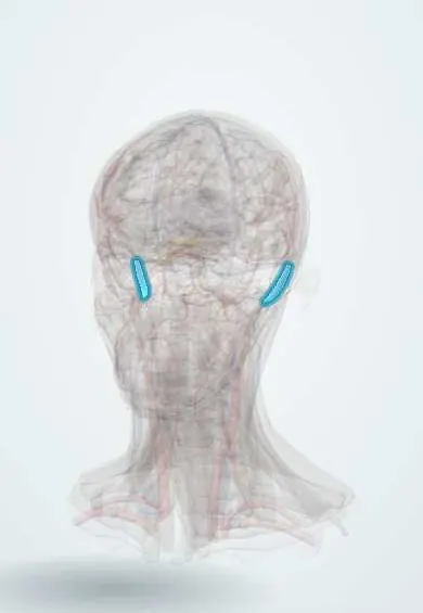
Cardiovascular system · Head and neck
Sigmoid sinus !
The sigmoid sinus is an S-shaped vein channel behind the ear that becomes the internal jugular vein; ear infections can spread to it.
Opens in the Durgical app, no sign-up needed.
What it is
An S-shaped sinus just behind the mastoid.
What it does
Becomes the internal jugular vein at the jugular foramen; ear infections can spread to it.
Key facts
- Drains
- Transverse and superior petrosal sinuses
- Into
- Internal jugular vein at the jugular foramen
Relations
- Course
- From the end of the transverse sinus at the petrous temporal bone it turns down and medially in the groove behind the mastoid, then forward onto the occipital bone to reach the back of the jugular foramen
- Lateral
- Mastoid cells and the mastoid process; the mastoid emissary vein leaves through the bone to the scalp veins behind the ear
- Medial
- Cerebellar hemisphere, across the dura
- In front
- In the jugular foramen, the glossopharyngeal nerve, vagus nerve and accessory nerve and the inferior petrosal sinus pass in front of it
How to find it
Lies behind the external ear canal, beneath the mastoid. In mastoid surgery its bluish wall bounds the dissection, and the retrosigmoid approach to the cerebellopontine angle is made just behind it.
Examination
- Griesinger sign: swelling and tenderness over the back of the mastoid from thrombosis of the mastoid emissary vein, pointing to sigmoid sinus thrombosis
On imaging
Contrast CT or MR venography shows thrombosis as a filling defect, sometimes with an empty delta sign; temporal bone CT shows opacified mastoid cells and erosion of the bony plate over the sinus.
Clinical notes
- Otogenic sinus thrombosis can extend into the internal jugular vein and shed septic emboli to the lungs
- Abscess beside the sinus (perisinus) and cerebellar abscess are other intracranial complications of mastoiditis
- A diverticulum or a gap in the bony wall of the sinus can cause pulsatile tinnitus
Anatomical variants
- A sinus lying unusually far forward, or a high jugular bulb, narrows the working space in ear surgery
Development
It is the lower end of the channel that forms above and behind the developing inner ear to bypass the closed part of the primary head vein; it joins the surviving lower part of that vein, the internal jugular.
Related conditions
Test yourself
Think of the answer, then tap the question to see it.
What separates the sigmoid sinus from the mastoid air cells?
Only a thin plate of bone, as it curves down in an S-shaped groove on the inner surface of the mastoid part of the temporal bone.
Which ear infection complication involves the sigmoid sinus, and how does it present?
Sigmoid (lateral) sinus thrombophlebitis from mastoiditis or middle-ear infection: spiking fevers and raised intracranial pressure (otitic hydrocephalus).
What is the Griesinger sign and what does it suggest?
Swelling and tenderness over the back of the mastoid from thrombosis of the mastoid emissary vein; it points to sigmoid sinus thrombosis.
Where does the sigmoid sinus end?
At the jugular foramen, where it continues as the internal jugular vein through the jugular bulb.
Educational information, not medical advice. For any symptom, see a health professional.
Its names
- Official Latin (TA2)
- sinus sigmoideus TA2 4847
- Official Spanish (DeCS)
- Seno sigmoideo DeCS D003392
- English (TA2)
- Sigmoid sinus
- Also called
- sinus sigmoides
Related
Sources: Terminologia Anatomica (TA2); DeCS; Neuroanatomy, Dural Venous Sinuses; Anatomy of the Human Body (20th ed.): Development of the Vascular System (Lea & Febiger (Gray's Anatomy, 1918), 1918) · adapted, Public domain.
Spotted a mistake?We are always improving for the community. Tell us what is wrong and our team will review it.Report a mistake
Representative model, for educational purposes only.