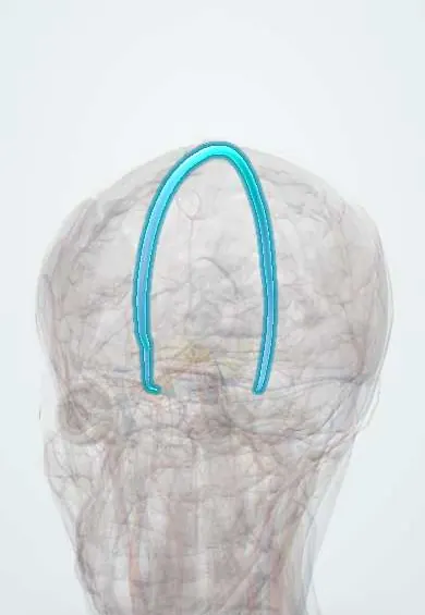
Cerebral venous sinus thrombosis !
A blood clot in the veins that drain the brain. It mostly affects young women, often around pregnancy or with the pill, and causes headache and seizures.
Updated 2026-11-01
Structures involved
Tap one to see what it is and open it in 3D.
- Superior sagittal sinus
- Transverse sinus
- Sigmoid sinus
- Straight sinus
- Confluence of sinuses
- Inferior sagittal sinus
- Cavernous sinus
- Internal jugular vein
- Optic disc
- White matter
Why it happens
- Hypercoagulable states: inherited and acquired
- Thrombosis and Virchow's triad
- The coagulation cascade
- Fibrinolysis and natural anticoagulants
What it is
Thrombosis of the dural venous sinuses and/or cerebral veins, which blocks venous drainage of the brain and raises intracranial pressure. It is an uncommon type of stroke that mainly affects young adults and women.
Who gets it
About 1–1.5 cases per 100,000 people per year in recent studies, around 0.5–1% of all strokes. Most patients are young adults, and women predominate (about 70%) because of oral contraceptives, pregnancy and the puerperium. In Latin America, pregnancy and the postpartum period are an important cause, so it is part of the assessment of headache in pregnant and postpartum women.
Causes
- Hormonal states: combined oral contraceptives, pregnancy and especially the puerperium, hormone therapy
- Inherited thrombophilias (factor V Leiden, prothrombin mutation, protein C, S or antithrombin deficiency) and antiphospholipid syndrome
- Local infection: otitis media, mastoiditis, sinusitis, meningitis, facial infections (septic thrombosis)
- Cancer, nephrotic syndrome, inflammatory diseases (lupus, Behçet disease, inflammatory bowel disease)
- Head trauma, neurosurgery or lumbar puncture; dehydration, especially in children
Risk factors
- Female sex, age 20 to 50
- Combined oral contraceptive use, especially with obesity
- Pregnancy and the first weeks postpartum
- Personal or family history of venous thrombosis
- Severe anaemia, polycythaemia and other blood disorders
Pathophysiology
A clot in a sinus (most often the superior sagittal and transverse sinuses) blocks venous outflow. Venous pressure rises, causing vasogenic and cytotoxic oedema and venous infarcts that are often haemorrhagic and do not follow an arterial territory. Blocked sinuses also absorb less CSF through the arachnoid granulations, so intracranial pressure rises and papilloedema appears.
Classification
- By location: superior sagittal sinus, transverse and sigmoid sinuses, deep system (straight sinus, internal cerebral veins), cortical veins, cavernous sinus
- By onset: acute (under 48 hours), subacute or chronic
- Septic (from nearby infection) or aseptic
Symptoms and signs
- Headache (in about 80–90%), usually progressive over days but sometimes sudden (thunderclap)
- Seizures, focal or generalised, in about 40%
- Focal deficits: weakness, aphasia, visual loss
- Signs of raised intracranial pressure: papilloedema, vomiting, double vision
- Drowsiness, confusion or coma, especially with deep venous thrombosis
Diagnosis
- Clinical
- Suspected in a young person, especially a woman, with an unusual or progressive headache plus seizures, focal signs or papilloedema, mainly when risk factors are present. Look for an infectious source in the ear, sinuses or face.
- Tests
- D-dimer is usually raised but a normal value does not exclude it. Blood count, coagulation, pregnancy test and a thrombophilia work-up (including antiphospholipid antibodies) at the right time; inflammatory markers if infection is suspected.
- Imaging
- MRI with MR venography or CT venography confirms the diagnosis by showing the clot and absent flow in the sinus. Non-contrast CT may be normal or show a hyperdense sinus (cord sign), an empty delta sign on contrast CT, or haemorrhagic venous infarcts.
Differential diagnosis
- Arterial ischaemic stroke
- Subarachnoid haemorrhage and other causes of thunderclap headache
- Idiopathic intracranial hypertension
- Brain tumour or abscess
- Preeclampsia or eclampsia with posterior reversible encephalopathy (in pregnancy)
Goal of treatment
Stop the clot from growing, help recanalisation and prevent new thrombosis, while controlling seizures and intracranial pressure and treating the underlying cause.
Treatment
- Medical
- Anticoagulation with heparin (low-molecular-weight heparin is usually preferred, unfractionated heparin when surgery may be needed) is the mainstay, even when there is haemorrhagic infarction.
- Medical
- After the acute phase, oral anticoagulation (vitamin K antagonists or, increasingly, direct oral anticoagulants) for a period that depends on the cause, usually months; longer when a persistent risk factor exists.
- Acute
- Antiseizure drugs for seizures, treatment of raised intracranial pressure, antibiotics and drainage of the source in septic thrombosis; stopping oestrogen-containing contraceptives.
- Procedure
- Endovascular thrombectomy or local thrombolysis in specialist centres for patients who worsen despite anticoagulation.
- Surgical
- Decompressive craniectomy for large lesions with impending herniation.
- Follow-up
- Neurological checks and level of consciousness, seizure watch, coagulation monitoring and bleeding precautions; follow-up imaging to check recanalisation.
Complications
- Haemorrhagic venous infarction and intracerebral haemorrhage
- Brain herniation, the main cause of early death
- Epilepsy after the event
- Visual loss from prolonged papilloedema
- Recurrent venous thrombosis, in the brain or elsewhere
Prognosis
Better than arterial stroke: with timely anticoagulation most patients recover independence, and death occurs in a small minority. Worse outcome is linked to coma, deep venous involvement, haemorrhage at admission, CNS infection and cancer. Residual headache, fatigue or cognitive problems are common.
Test yourself
Think of the answer, then tap the question to see it.
Which patient profile should raise suspicion of cerebral venous thrombosis?
A young woman on oral contraceptives, pregnant or postpartum, with progressive headache, seizures, focal signs or papilloedema.
Which imaging study confirms cerebral venous thrombosis?
MRI with MR venography, or CT venography, showing the clot and absent flow in the venous sinus.
Is anticoagulation contraindicated in cerebral venous thrombosis with haemorrhagic infarction?
No. Heparin anticoagulation is the mainstay even when venous haemorrhage is present.
Which dural venous sinus is most often involved in cerebral venous thrombosis?
The superior sagittal sinus, followed by the transverse sinuses.
What is the classic sign of superior sagittal sinus thrombosis on contrast CT?
The empty delta sign: a filling defect surrounded by enhancing sinus walls.
Related conditions
- Idiopathic intracranial hypertension
- Ischaemic stroke
- Intracerebral haemorrhage
- Subarachnoid haemorrhage
- Inherited thrombophilia
- Antiphospholipid syndrome
- Preeclampsia
- Bacterial meningitis
- Acute otitis media
- Epilepsy and seizures
- Systemic lupus erythematosus
Educational information, not medical advice. For any symptom, see a health professional.
Sources: Diagnosis and Treatment of Cerebral Venous Thrombosis: A Review (Frontiers in Aging Neuroscience, 2018).
Spotted a mistake?We are always improving for the community. Tell us what is wrong and our team will review it.Report a mistake
Representative model, for educational purposes only.