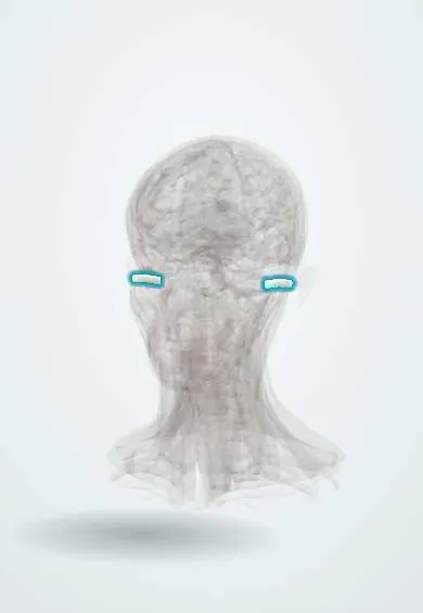
Ear canal !
The ear canal is the S-shaped tube, about 2.5 cm long, from the outer ear to the eardrum; it carries sound inward and makes earwax.
Official name: External acoustic meatus
Opens in the Durgical app, no sign-up needed.
What it is
The 2.5 cm S-shaped canal from the outer ear to the eardrum.
What it does
Carries sound to the eardrum; it makes wax and can get infected (swimmer's ear).
Key facts
- Where
- From the concha to the tympanic membrane (~2.5 cm, S-shaped)
- Blood supply
- Posterior auricular, maxillary, superficial temporal arteries
- Nerve
- V3 (auriculotemporal), X (Arnold's nerve: cough when touched); great auricular (C2–C3) to the back; small VII twig to the posterior wall
- Function
- Conducts sound; makes wax
Relations
- Medial
- Tympanic membrane, set obliquely so the anteroinferior canal wall is longer and forms a recess where debris collects
- In front
- Head of the mandible at the temporomandibular joint: jaw movement is felt with a finger in the canal, and a blow to the chin can fracture the anterior bony wall
- Behind
- Mastoid air cells; in mastoiditis the posterosuperior canal wall may sag
- Above
- Thin bone separating it from the middle cranial fossa and the temporal lobe
- Below
- Parotid gland; small gaps in the floor of the cartilage (fissures of Santorini) let infection or tumour pass between canal and parotid
How to find it
With the auricle drawn up and back, the otoscope speculum is passed just into the cartilaginous part (the bony part is very sensitive) and the eardrum is seen at the end, facing slightly down and forward. Wax normally migrates outward on its own with the skin of the canal.
Examination
- Tragal pressure or auricle traction causing sharp pain: otitis externa
- Otoscopy: a swollen, narrowed canal with debris (otitis externa), fluffy white or black material (fungal infection), wax plug, foreign body or bony swellings
- Rinne and Weber tests: wax or a foreign body blocking the canal produces a conductive pattern (Rinne negative, Weber to the blocked side)
On imaging
Seen directly with the otoscope. High-resolution temporal bone CT shows its bony part: narrowing or absence (atresia), exostoses (broad, often both sides, in cold-water swimmers), a single stalked osteoma, and bone erosion. In necrotising (malignant) otitis externa, CT shows erosion of the canal floor and skull base, and MRI shows soft tissue spread better.
Clinical notes
- Otitis externa (swimmer's ear) follows moisture, scratching or cotton-bud trauma that breaks the skin and its waxy barrier; Pseudomonas aeruginosa and Staphylococcus aureus are the usual bacteria
- Foreign bodies are common in young children; insects and button batteries are urgent, the latter because they burn the canal skin quickly
- Exostoses (surfer's ear): smooth bony swellings of the deep canal after repeated cold-water exposure; large ones trap water and wax
- Necrotising otitis externa can involve the facial nerve at the stylomastoid foramen and lower cranial nerves at the skull base
Development
The canal comes from the first pharyngeal cleft; a solid meatal plug later hollows out, and failure produces congenital aural atresia, often with a malformed auricle.
Related conditions
Test yourself
Think of the answer, then tap the question to see it.
How do the lateral and medial parts of the external acoustic meatus differ?
Lateral third: cartilaginous, with hairs and glands; medial two thirds: bony, thin skin without glands. Furuncles arise only in the outer part.
How is the ear canal straightened for otoscopy in adults and in infants?
Adults: auricle pulled up and back; infants: down and back, as their canal is short and mostly cartilaginous.
What is necrotising (malignant) otitis externa?
Severe canal infection with granulation tissue in the floor, in an older person with diabetes or immunosuppression; it spreads to the skull base.
Which nerve explains a cough when the ear canal is touched?
The auricular branch of the vagus nerve (Arnold nerve), which supplies part of the external acoustic meatus.
Educational information, not medical advice. For any symptom, see a health professional.
Its names
- Official Latin (TA2)
- meatus acusticus externus TA2 6867
- Official Spanish (DeCS)
- Conducto Auditivo Externo DeCS D004424
- English (TA2)
- External acoustic meatus
Related
Sources: Terminologia Anatomica (TA2); DeCS; Ear, Hearing, and Facial Nerve Evaluation; Otitis Externa.
Spotted a mistake?We are always improving for the community. Tell us what is wrong and our team will review it.Report a mistake
Representative model, for educational purposes only.