Cardiovascular system · Head and neck
Petrosal sinuses !
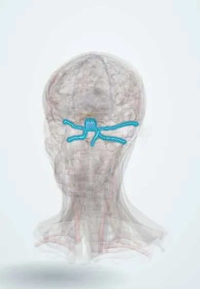
The petrosal sinuses are small vein channels along the skull base, near the ear bone, that drain the cavernous sinus toward the neck veins.
Official name: Superior petrosal sinus
Opens in the Durgical app, no sign-up needed.
What it is
Smaller sinuses along the petrous bone and the back of the skull base.
What it does
Drain the cavernous sinus and the skull base.
Key facts
- Drains
- Cavernous sinus, skull base
- Into
- Superior -> transverse/sigmoid; inferior -> internal jugular
Relations
- Course
- Both leave the back of the cavernous sinus: the superior runs back along the petrous ridge to the transverse–sigmoid junction; the inferior runs down and back to the jugular foramen
- Above
- Above the superior sinus: the attached edge of the tentorium cerebelli and the temporal lobe
- Below
- The trigeminal nerve passes beneath the superior sinus at the petrous apex into Meckel's cave; the abducens nerve runs close to the inferior sinus in Dorello canal
- Medial
- Clivus with the basilar venous plexus, which links the two sides
How to find it
Neither can be felt. The superior sinus is seen along the petrous ridge in the retrosigmoid approach, where the petrosal vein joins it; the inferior sinus is reached by a catheter passed from the femoral vein up the internal jugular vein for sampling.
On imaging
MR or CT venography shows the superior sinus along the petrous ridge and the inferior sinus in the groove between the petrous bone and the clivus. In ACTH-dependent Cushing syndrome, inferior petrosal sinus sampling by catheter compares ACTH from both sinuses with peripheral blood to confirm a pituitary source. They also take part in cavernous sinus thrombosis and dural fistulas.
Clinical notes
- Petrous apex infection from otitis media can affect the abducens nerve nearby: Gradenigo syndrome (ear discharge, pain around the eye, lateral rectus palsy)
- The petrosal vein (of Dandy) drains the cerebellum and brainstem into the superior petrosal sinus; it is met in cerebellopontine angle surgery such as microvascular decompression for trigeminal neuralgia
- Thrombosis of the cavernous sinus can spread along them toward the sigmoid sinus and internal jugular vein
Anatomical variants
- The inferior petrosal sinus joins the internal jugular vein in varying ways, sometimes outside the skull or into the vertebral venous plexus, which can make catheter sampling difficult
Development
The superior petrosal sinus is the old drainage trunk of the middle embryonic dural plexus, which turns to carry blood from the cavernous sinus to the transverse sinus; the inferior petrosal sinus appears later.
Test yourself
Think of the answer, then tap the question to see it.
Where do the superior and inferior petrosal sinuses drain the cavernous sinus to?
The superior petrosal sinus to the junction of the transverse and sigmoid sinuses; the inferior petrosal sinus to the internal jugular vein through the jugular foramen.
What is inferior petrosal sinus sampling used for?
Blood from both sides is tested for pituitary ACTH to confirm Cushing disease and suggest the tumour's side.
What is Gradenigo syndrome?
Petrous apex infection from otitis media affecting the nearby abducens nerve: ear discharge, pain around the eye and lateral rectus palsy.
Educational information, not medical advice. For any symptom, see a health professional.
Its names
- Official Latin (TA2)
- sinus petrosus superior TA2 4850
- Official Spanish (DeCS)
- Senos petrosos y occipital DeCS D003392
- English (TA2)
- Superior petrosal sinus
Sources: Terminologia Anatomica (TA2); DeCS; Neuroanatomy, Superior Petrosal Sinus; Neuroanatomy, Dural Venous Sinuses; Anatomy of the Human Body (20th ed.): Development of the Vascular System (Lea & Febiger (Gray's Anatomy, 1918), 1918) · adapted, Public domain.
Spotted a mistake?We are always improving for the community. Tell us what is wrong and our team will review it.Report a mistake
Representative model, for educational purposes only.