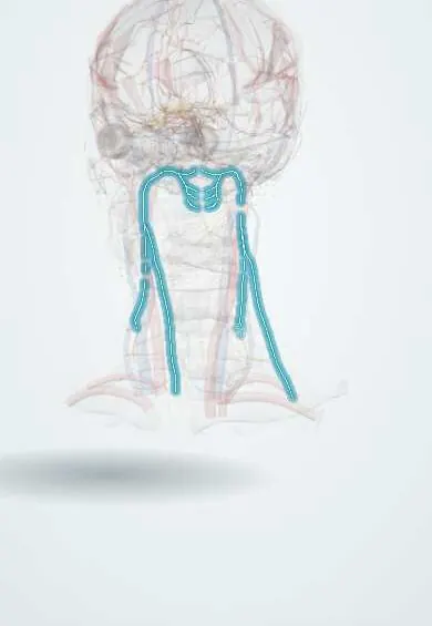
Nervous system · Head and neck
Accessory nerve (XI) !
The accessory nerve (cranial nerve XI) moves the sternocleidomastoid and trapezius muscles, which turn the head and shrug the shoulders.
Official name: Accessory nerve
Opens in the Durgical app, no sign-up needed.
What it is
A nerve that runs across the back triangle of the neck to the trapezius.
What it does
Moves the sternocleidomastoid and trapezius (shrugging); easily injured in neck biopsies.
Key facts
- Roots
- Spinal accessory nucleus (C1–C5 cord); rises through the foramen magnum, exits the jugular foramen; classic texts also describe a cranial root (caudal nucleus ambiguus) that joins the vagus as the internal branch and supplies the larynx
- Moves
- Sternocleidomastoid, trapezius
- Feeling from
- None
Relations
- Course
- Rootlets from the upper cervical spinal cord join and rise through the foramen magnum, then leave through the jugular foramen with the vagus nerve (X); the nerve passes in front of the internal jugular vein, supplies and usually pierces the sternocleidomastoid, crosses the posterior triangle on the levator scapulae and reaches the anterior border of the upper trapezius
- Deep
- In the posterior triangle, the levator scapulae and the splenius in its floor
- Superficial
- Only the investing layer of deep cervical fascia and the skin; superficial neck lymph nodes lie along it
How to find it
Its surface line runs from about a third of the way down the posterior border of the sternocleidomastoid, across the posterior triangle, to the anterior border of the trapezius in the lower neck; lymph nodes along this line are where it is at risk.
Examination
- Shoulder shrug against resistance: weakness on one side points to the trapezius supply
- Head turning against the examiner's hand on the chin: weakness turning to one side means a weak sternocleidomastoid on the opposite side (the left muscle turns the face to the right)
- Raising the arm above the head: shoulder droop and lateral winging of the scapula point to trapezius weakness
On imaging
Not seen on radiographs. High-resolution ultrasound can follow it across the posterior triangle just under the investing fascia, from the posterior border of the sternocleidomastoid, and show a cut nerve, neuroma or scar entrapment after node biopsy. Old injury leaves a thin trapezius on CT or MRI. For skull base tumours, MRI checks the jugular foramen, where it exits.
Clinical notes
- The injury can go unnoticed at first; shoulder aching and weakness appear in the following weeks as the trapezius wastes
- Modified radical neck dissection preserves it where possible, because sacrificing it leaves lasting shoulder dysfunction
- Jugular foramen lesions affect it together with IX and X (Vernet syndrome)
Anatomical variants
- It usually passes in front of the internal jugular vein at the top of the neck, less often behind it
- It may pass deep to the sternocleidomastoid instead of piercing it
Related conditions
Test yourself
Think of the answer, then tap the question to see it.
Where is the accessory nerve (XI) most often injured, and what is the result?
In posterior triangle surgery such as lymph node biopsy: trapezius weakness with a drooping shoulder, shoulder pain and difficulty raising the arm.
Why does an accessory nerve injury in the posterior triangle spare the sternocleidomastoid?
The branch to the sternocleidomastoid leaves higher up, so only the trapezius weakens.
How does scapular winging from accessory nerve palsy differ from long thoracic nerve palsy?
Trapezius palsy gives lateral winging on abduction; serratus anterior (long thoracic) palsy gives medial winging.
How is the accessory nerve tested at the bedside?
Shoulder shrug against resistance (trapezius) and head turning against the hand (sternocleidomastoid turns the face to the opposite side).
Educational information, not medical advice. For any symptom, see a health professional.
Its names
- Official Latin (TA2)
- nervus accessorius TA2 6352
- Official Spanish (DeCS)
- Nervio Accesorio DeCS D000055
- English (TA2)
- Accessory nerve
- Also called
- spinal accessory nerve · cranial nerve XI · nerve of Willis
Related
Sources: Terminologia Anatomica (TA2); DeCS; Neuroanatomy, Cranial Nerve 11 (Accessory).
Spotted a mistake?We are always improving for the community. Tell us what is wrong and our team will review it.Report a mistake
Representative model, for educational purposes only.