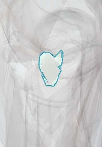
Gluteus minimus tendon !
The gluteus minimus tendon attaches to the front of the bony point of the outer hip (greater trochanter), lying right on the hip capsule.
Official name: Tendon of gluteus minimus
Opens in the Durgical app, no sign-up needed.
What it is
The tendon of the gluteus minimus, attaching to the front of the greater trochanter and partly to the hip capsule.
What it does
Carries the pull of the gluteus minimus to the femur to keep the pelvis level.
Key facts
- Attaches
- Anterior facet of the greater trochanter; some fibres to the superior hip capsule
- Function
- Abduction and medial rotation; with the gluteus medius keeps the pelvis level; torn together with the medius in trochanteric tears
- Tight in
- Hip adduction with lateral rotation
Relations
- Superficial
- Gluteus medius tendon, with the superior gluteal nerve and vessels and a small bursa between them
- Deep
- Upper hip capsule and the reflected head of the rectus femoris
- Behind
- Piriformis tendon
How to find it
Not felt separately; on ultrasound it is the tendon on the anterior facet, the most anterior of the greater trochanter's attachments.
Examination
- Trendelenburg test: in single-leg stance the pelvis drops on the opposite side when the abductors (medius and minimus together) are weak or torn
- Single-leg stance for 30 seconds: lateral hip pain within that time suggests gluteal tendinopathy, which often involves the minimus as well as the medius
On imaging
Ultrasound shows its insertion on the anterior facet; MRI shows tears, calcific tendinopathy and fatty change of the muscle.
Common problems
- Gluteal tendinopathy: wear and pain of its tendon at the outer hip, common in women over 40 and in runners. It is the main cause of greater trochanteric pain syndrome.
- Tendon tears, sometimes called a 'rotator cuff tear of the hip'.
- Weakness causes a limp where the pelvis drops on the other side (Trendelenburg gait).
How it is diagnosed
Physical exam: tenderness over the outer hip, pain when standing on one leg, and strength testing. Ultrasound or MRI shows tendon damage or tears.
Treatment
- Physical therapy with progressive loading exercises; avoiding crossing the legs and lying on the sore side.
- Injections or shock wave therapy for pain that does not settle.
- Surgical repair of the tendon (open or endoscopic) for significant tears.
Clinical notes
- Repairs of tears at the trochanter usually address the gluteus medius and minimus tendons together
- Anterolateral and direct lateral hip approaches split or partly detach the front of the minimus, which is repaired at closure; poor healing adds to a limp after surgery
- Isolated minimus tears are uncommon; on MRI they show as fluid or a gap at the anterior facet of the greater trochanter
Related conditions
Test yourself
Think of the answer, then tap the question to see it.
Where does the gluteus minimus tendon insert?
On the anterior facet of the greater trochanter, with some fibres to the superior hip capsule.
Which nerve runs between the gluteus minimus and the gluteus medius?
The superior gluteal nerve, with its vessels; injury to it weakens both muscles.
With which tendon is the gluteus minimus tendon usually torn?
The gluteus medius tendon; repairs at the trochanter usually address both together.
Educational information, not medical advice. For any symptom, see a health professional.
Its names
- Official Latin (TA2)
- musculus gluteus minimus TA2 2600
- Official Spanish (DeCS)
- Tendón del glúteo menor DeCS D013710
- English (TA2)
- Tendon of gluteus minimus
- Also called
- musculus glutaeus minimus
Related
Sources: Terminologia Anatomica (TA2); DeCS; Anatomy, Bony Pelvis and Lower Limb, Gluteus Minimus Muscle.
Spotted a mistake?We are always improving for the community. Tell us what is wrong and our team will review it.Report a mistake
Representative model, for educational purposes only.