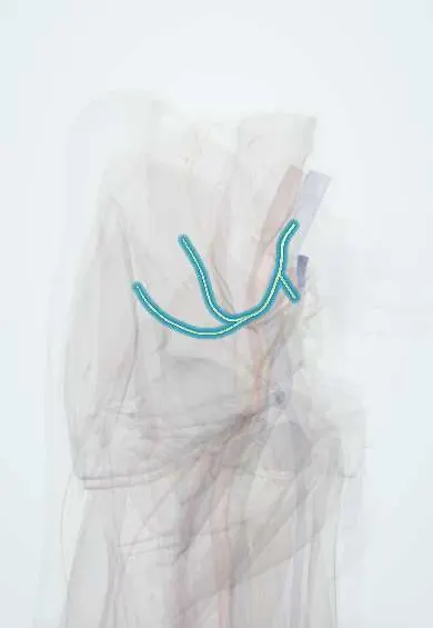
Superior gluteal nerve !
The superior gluteal nerve supplies the gluteus medius, gluteus minimus and tensor fasciae latae, which keep the pelvis level.
Opens in the Durgical app, no sign-up needed.
What it is
A nerve that leaves the pelvis above the piriformis and runs between the gluteus medius and minimus.
What it does
Controls the gluteus medius, gluteus minimus and TFL, which keep the pelvis level when walking.
Key facts
- Roots
- L4–S1
- Moves
- Gluteus medius, gluteus minimus, tensor fasciae latae; injury gives positive Trendelenburg sign
- Feeling from
- None to skin (no cutaneous branch)
Relations
- Course
- Leaves the pelvis through the greater sciatic foramen above the piriformis with the superior gluteal vessels, turns forward between the gluteus medius and gluteus minimus and divides into an upper branch along the upper border of the minimus and a lower branch across it to the tensor fasciae latae
- Superficial
- The gluteus medius, with the gluteus maximus over it
- Deep
- The gluteus minimus; at its exit, the bone of the greater sciatic notch
- Below
- The piriformis, which separates it from the sciatic nerve, the inferior gluteal nerve and the posterior femoral cutaneous nerve below
How to find it
It cannot be felt. Its exit from the pelvis, with the superior gluteal artery, lies at the junction of the upper and middle thirds of a line from the posterior superior iliac spine to the tip of the greater trochanter.
Examination
- Trendelenburg test: standing on the affected leg, the opposite side of the pelvis drops when the hip abductors or their nerve fail; it is also positive in a painful or dislocated hip and in coxa vara
- Resisted hip abduction lying on the side: weakness with normal knee and ankle strength points to this nerve; weakness together with foot drop suggests an L5 root lesion
On imaging
Too small to see reliably on routine imaging. Its exit from the pelvis above the piriformis, with the superior gluteal artery, is the landmark on MRI or CT. A lesion shows indirectly: denervation oedema and later fatty atrophy of the gluteus medius, gluteus minimus and tensor fasciae latae. MR neurography may show the nerve itself. Radiographs do not show it.
Common problems
- Injury, for example in some hip surgery approaches, weakens these muscles and causes a limp.
Clinical notes
- At risk in the direct lateral (transgluteal) hip approach and, for its branch to the tensor fasciae latae, in anterolateral approaches
- Intramuscular injections placed too close to the greater sciatic notch can reach it
- Bilateral weakness of the abductors produces a waddling gait; on one side, the trunk leans over the stance leg to compensate
Anatomical variants
- It usually divides into two branches (about 6 in 7 people); the rest have three
Test yourself
Think of the answer, then tap the question to see it.
What makes the superior gluteal nerve unique among nerves leaving the pelvis through the greater sciatic foramen?
It is the only nerve to leave above the piriformis, together with the superior gluteal artery and vein.
Which muscles does the superior gluteal nerve supply?
Gluteus medius, gluteus minimus and tensor fasciae latae; it has no skin branch.
Which sign follows a superior gluteal nerve lesion?
A positive Trendelenburg sign: in single-leg stance on the injured side, the pelvis drops on the opposite side.
How does resisted hip abduction help separate a superior gluteal nerve lesion from an L5 root lesion?
Abduction weakness with normal knee and ankle strength points to the nerve; weakness together with foot drop suggests L5.
Educational information, not medical advice. For any symptom, see a health professional.
Its names
- Official Latin (TA2)
- nervus gluteus superior TA2 6543
- Official Spanish (DeCS)
- Nervio glúteo superior
- English (TA2)
- Superior gluteal nerve
- Also called
- nervus glutaeus superior · nervus glutaeus cranialis
Related
Sources: Terminologia Anatomica (TA2); Anatomy, Abdomen and Pelvis: Superior Gluteal Nerve.
Spotted a mistake?We are always improving for the community. Tell us what is wrong and our team will review it.Report a mistake
Representative model, for educational purposes only.