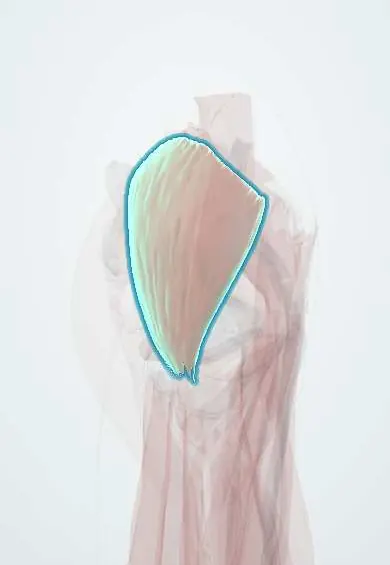
Gluteus medius !
The gluteus medius is a muscle on the side of the hip; it keeps the pelvis level when standing on one leg.
Official name: Gluteus medius muscle
Opens in the Durgical app, no sign-up needed.
What it is
A fan-shaped muscle on the outer side of the hip, partly under the gluteus maximus. It runs from the outer pelvis to the greater trochanter, the bony point of the outer hip.
What it does
Moves the leg out to the side and, most importantly, keeps the pelvis level when you stand on one leg, as happens with every step.
Key facts
- Origin
- External ilium between anterior and posterior gluteal lines
- Insertion
- Lateral surface of greater trochanter
- Action
- Abducts and medially rotates hip; keeps pelvis level in single-leg stance (weakness: Trendelenburg sign)
- Nerve
- Superior gluteal nerve (L4–S1)
- Blood supply
- Superior gluteal artery (deep branch)
Relations
- Superficial
- Gluteus maximus over its back part; the front part is covered only by the strong gluteal aponeurosis
- Deep
- Gluteus minimus, with the superior gluteal nerve and the deep branch of the superior gluteal artery running between them
- In front
- Tensor fasciae latae
- Below
- Piriformis along its lower border; the superior gluteal vessels and nerve emerge between the two
- Lateral
- Trochanteric bursa between its tendon and the gluteus maximus and iliotibial tract; a smaller bursa lies deep to its own tendon
How to find it
Lying on one side with the upper leg lifted against resistance, the muscle is felt contracting below the iliac crest and above the greater trochanter. Standing on one leg, it tightens under the fingers on the stance side.
Examination
- Trendelenburg test: standing on the affected leg, the pelvis drops on the opposite side (or the trunk leans over the stance side); this points to weak hip abductors, a superior gluteal nerve lesion or a mechanical hip problem
- Single-leg stance for 30 seconds: reproduction of outer hip pain supports gluteal tendinopathy
- Resisted abduction lying on the side: weakness or pain over the greater trochanter suggests tendinopathy or a tendon tear
On imaging
Ultrasound and MRI of the greater trochanter are first line for lateral hip pain. The tendon inserts on the lateral and superoposterior facets. Tendinopathy is a thick tendon; a tear is a gap or detachment, often with trochanteric bursitis. Late fatty atrophy on MRI predicts a poor repair. Radiographs show only calcification or an irregular trochanter.
Common problems
- Gluteal tendinopathy: wear and pain of its tendon at the outer hip, common in women over 40 and in runners. It is the main cause of greater trochanteric pain syndrome.
- Tendon tears, sometimes called a 'rotator cuff tear of the hip'.
- Weakness causes a limp where the pelvis drops on the other side (Trendelenburg gait).
How it is diagnosed
Physical exam: tenderness over the outer hip, pain when standing on one leg, and strength testing. Ultrasound or MRI shows tendon damage or tears.
Treatment
- Physical therapy with progressive loading exercises; avoiding crossing the legs and lying on the sore side.
- Injections or shock wave therapy for pain that does not settle.
- Surgical repair of the tendon (open or endoscopic) for significant tears.
Clinical notes
- Superior gluteal nerve injury after a lateral or anterior hip approach, or from a badly placed injection, causes a Trendelenburg gait
- Tears of the abductor tendons are found on MRI or during hip arthroplasty in older people with long-standing lateral hip pain; some are repaired
- In the lateral (transgluteal) approach, the front part of the muscle is split or detached and then repaired; failure of the repair leaves a lasting limp
Development
A dorsal-mass muscle, as its nerve shows: the superior gluteal nerve (L4–S1) arises from posterior divisions of the sacral plexus and is shared with the gluteus minimus and the tensor fasciae latae.
Related conditions
Test yourself
Think of the answer, then tap the question to see it.
What is the nerve supply of the gluteus medius?
The superior gluteal nerve (L4–S1).
What is the key function of the gluteus medius during walking?
Keeping the pelvis level in single-leg stance; it also abducts and medially rotates the hip.
What does a positive Trendelenburg test show with gluteus medius weakness?
Standing on the affected leg, the pelvis drops on the opposite side, or the trunk leans over the stance side to compensate.
Why is the gluteus medius not split far above the greater trochanter in a lateral hip approach?
The superior gluteal nerve runs between gluteus medius and minimus about 5 cm above the trochanter tip.
How is the ventrogluteal injection site over the gluteus medius located?
Palm on the greater trochanter, index finger on the ASIS, middle finger along the iliac crest; the V between them lies far from the sciatic nerve.
Educational information, not medical advice. For any symptom, see a health professional.
Its names
- Official Latin (TA2)
- musculus gluteus medius TA2 2599
- Official Spanish (DeCS)
- Músculo glúteo medio
- English (TA2)
- Gluteus medius muscle
- Also called
- musculus glutaeus medius
Related
- Gluteus maximus
- Gluteus minimus
- Tensor fasciae latae
- Trochanteric bursae
- Piriformis
- Superior gluteal nerve
- Gluteal arteries
- Gluteus medius tendon
Sources: Terminologia Anatomica (TA2); Anatomy, Bony Pelvis and Lower Limb, Gluteus Medius Muscle.
Spotted a mistake?We are always improving for the community. Tell us what is wrong and our team will review it.Report a mistake
Representative model, for educational purposes only.