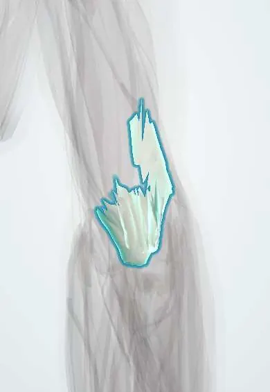
Triceps tendon !
The triceps tendon attaches the triceps to the point of the elbow (olecranon); it straightens the elbow and only rarely ruptures.
Opens in the Durgical app, no sign-up needed.
What it is
The broad tendon at the back of the elbow joining the three triceps heads to the olecranon.
What it does
Straightens the elbow.
Key facts
- Attaches
- Common tendon of the three heads to the upper surface of the olecranon; lateral expansion to the fascia over the anconeus
- Function
- Elbow extension; the olecranon bursa lies over it; rupture is rare, after a fall on the outstretched hand
- Tight in
- Elbow flexion with shoulder flexion
Relations
- Course
- The three heads converge on the back of the lower arm into a superficial layer (long and lateral heads) and a deeper medial-head part, which insert together on the olecranon
- Superficial
- Skin and the subcutaneous olecranon bursa over the tip of the olecranon
- Deep
- Elbow capsule and the olecranon fossa; a small triceps bursa lies between the tendon and the olecranon
- Medial
- Ulnar nerve in the cubital tunnel, behind the medial epicondyle
- Lateral
- Anconeus, into whose fascia its lateral expansion blends
How to find it
With the arm forward and the elbow bent, the tendon is felt as a broad band just above the olecranon. A palpable gap there after a fall, with weak extension, suggests a rupture.
Examination
- Modified Thompson test: arm supported with the elbow bent over the edge of a table; squeezing the triceps belly produces no extension when the tendon is completely torn
- Resisted extension and extension overhead against gravity: weakness with a gap suggests a tear; a partial tear keeps some extension
- Triceps reflex (C7): tapping the tendon just above the olecranon contracts the muscle; loss suggests a C7 root or radial nerve lesion
On imaging
A lateral elbow X-ray may show a bony flake just above the olecranon (fleck sign) after an avulsion. Ultrasound and sagittal MRI separate partial from complete tears, show which layer is torn and how far it has retracted.
Clinical notes
- Posterior approaches to the elbow for distal humerus fractures split, reflect or work beside the tendon, or use an olecranon osteotomy; it is repaired securely afterward
- Snapping triceps: the medial head can slip over the medial epicondyle, sometimes with the ulnar nerve, causing medial snapping
- Its pull separates displaced olecranon fracture fragments, which is why they need fixation (for example a tension band)
Anatomical variants
- A rare fourth head can occur and may compress the radial or ulnar nerve
Development
The olecranon has its own apophysis, which appears late in childhood and fuses in mid-adolescence. Before fusion the triceps tends to avulse the apophysis rather than tear, and a physis that stays open can be mistaken for a fracture; comparing with the other elbow helps.
Test yourself
Think of the answer, then tap the question to see it.
Where does the triceps tendon insert?
On the upper surface of the olecranon, with a lateral expansion to the fascia over the anconeus.
What X-ray sign suggests a triceps tendon rupture?
The fleck sign: a small flake of olecranon pulled off, seen on a lateral X-ray.
Why is a modified Thompson test used for triceps tendon rupture?
Extension against gravity may survive a complete tear through the lateral expansion and anconeus; squeezing the belly gives no extension if torn.
Which nerve root does the triceps reflex test?
C7: tapping the triceps tendon just above the olecranon contracts the muscle; loss suggests a C7 root or radial nerve lesion.
Educational information, not medical advice. For any symptom, see a health professional.
Its names
- Official Latin (TA2)
- tendo musculi tricipitis brachii TA2 2471
- Official Spanish (DeCS)
- Tendón del músculo tríceps braquial
- English (TA2)
- Triceps tendon
Related
- Olecranon bursa
- Triceps bursa
- Humerus
- Ulna
- Triceps (long head)
- Triceps (lateral head)
- Triceps (medial head)
Sources: Terminologia Anatomica (TA2); Anatomy, Shoulder and Upper Limb, Triceps Muscle.
Spotted a mistake?We are always improving for the community. Tell us what is wrong and our team will review it.Report a mistake
Representative model, for educational purposes only.