Joints and ligaments · Upper limb
Elbow capsule !
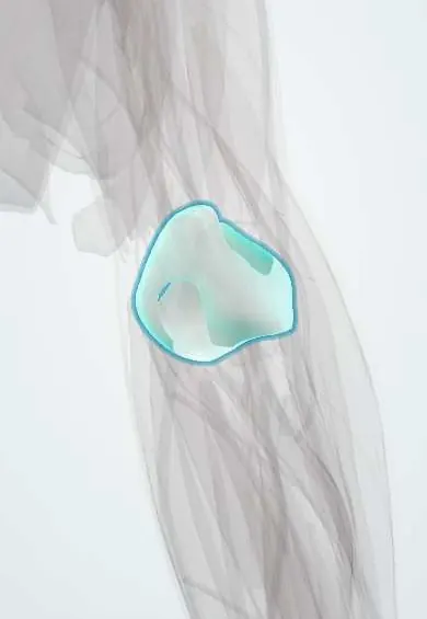
The elbow capsule is the sleeve that seals the elbow joint; after an injury it can stiffen and stop the elbow straightening fully.
Official name: Elbow joint capsule
Opens in the Durgical app, no sign-up needed.
What it is
The thin bag of tissue around the elbow joint.
What it does
Seals the joint; a stiff capsule after injury limits straightening.
Key facts
- Attaches
- Humerus above the coronoid, radial and olecranon fossae to the coronoid margin and annular ligament
- Function
- Encloses the humeroulnar, humeroradial and proximal radioulnar joints; thickened by the collateral ligaments; flexion–extension arc about 140–145°
Relations
- In front
- Brachialis, partly attached to it; in the cubital fossa in front, the brachial artery and median nerve
- Behind
- Triceps tendon and anconeus, with the posterior fat pad between capsule and bone in the olecranon fossa
- Medial
- Thickened by the ulnar collateral ligament (UCL); the ulnar nerve lies against the posteromedial capsule behind the medial epicondyle
- Lateral
- Thickened by the radial collateral ligament and blended with the annular ligament; the radial nerve divides in front of it, and its posterior interosseous branch enters the supinator
How to find it
With the elbow bent at 90°, the soft spot is felt in the triangle between the lateral epicondyle, the radial head (which turns under the thumb as the forearm rotates) and the olecranon tip; a bulge here means an effusion.
Examination
- Elbow extension test: after a fall, full active extension makes a fracture needing treatment unlikely; inability to straighten the elbow fully suggests an effusion or fracture and calls for X-rays
On imaging
Lateral X-ray at 90° flexion: a raised anterior fat pad forms a sail sign, and any visible posterior fat pad is abnormal. In children this suggests a supracondylar fracture, in adults a radial head fracture, even with no fracture line. Ultrasound shows fluid distending the posterior recess.
Clinical notes
- Septic arthritis of the elbow: painful, swollen joint held flexed; fluid is aspirated through the soft spot for cell count, Gram stain, culture and crystals
- Post-traumatic stiffness that does not respond to therapy is treated by open or arthroscopic capsular release; a functional arc of about 30–130° of flexion is the commonly quoted goal
- Arthroscopy portals sit close to nerves (the radial nerve in front laterally, the ulnar nerve medially); the joint is distended with fluid first to push them away
Development
The joint cavity appears in the embryo when the central interzone tissue between the cartilage models dies back, the outer mesenchyme forming the capsule; in children, the ossification centres that appear inside it in a fixed order (CRITOE) must be known to read elbow films.
Related conditions
Test yourself
Think of the answer, then tap the question to see it.
What does a visible posterior fat pad on a lateral elbow X-ray mean?
Fluid in the joint: the fat pads lie inside the elbow capsule but outside the synovium, and an effusion lifts them.
Where is the elbow joint aspirated?
Through the lateral soft spot, the centre of the triangle formed by the lateral epicondyle, radial head and olecranon tip.
Which movement is lost first when the elbow capsule stiffens after trauma?
The last degrees of extension, as the anterior capsule contracts.
Educational information, not medical advice. For any symptom, see a health professional.
Its names
- Official Latin (TA2)
- capsula articularis articulationis cubiti TA2 1536
- Official Spanish (DeCS)
- Cápsula articular de la articulación del codo
- English (TA2)
- Elbow joint capsule
Related
Sources: Terminologia Anatomica (TA2); Anatomy, Shoulder and Upper Limb, Elbow Joint; 9.7 Development of Joints (OpenStax (Rice University), 2022) · adapted, CC BY 4.0; Anatomía humana (4.ª ed.), tomo 1, cap. 56: Articulación del codo. Músculos del brazo.
Spotted a mistake?We are always improving for the community. Tell us what is wrong and our team will review it.Report a mistake
Representative model, for educational purposes only.