Conditions · Pediatrics and neonatology
Esophageal atresia and tracheoesophageal fistula !
A birth defect in which the oesophagus ends in a blind pouch, often with an abnormal link to the windpipe. It needs surgery soon after birth.
Updated 2026-11-01
Structures involved
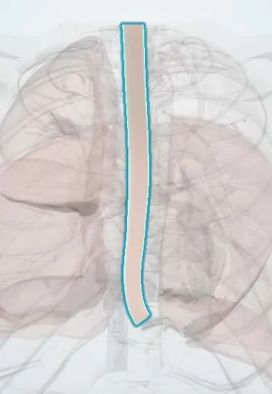
Tap one to see what it is and open it in 3D.
What it is
A congenital interruption of the oesophagus, which ends in a blind upper pouch, usually combined with an abnormal connection (fistula) between the trachea and the lower oesophagus. It prevents feeding and puts the lungs at risk of aspiration.
Who gets it
Occurs in about 1 in 3,000 to 4,500 live births. Roughly half of affected babies have other anomalies, especially cardiac and those of the VACTERL association. Prenatal ultrasound and early neonatal surgery are available in Colombian referral centres, but late diagnosis still happens where prenatal care is limited.
Causes
- Abnormal separation of the foregut into trachea and oesophagus in the 4th to 6th week of gestation
- Usually sporadic, with a multifactorial cause
- Associated with chromosomal anomalies (trisomy 18, trisomy 21) and syndromes such as VACTERL and CHARGE
Risk factors
- Polyhydramnios in pregnancy (the fetus cannot swallow amniotic fluid)
- Other congenital anomalies, especially heart defects
- Prematurity and low birth weight (associated, not causal)
Pathophysiology
The upper oesophagus ends blindly, so saliva and feeds pool and overflow into the airway. In the most common type, the lower oesophagus connects to the trachea, letting air enter the stomach and gastric acid reflux into the lungs. The trachea wall at the fistula site is often weak (tracheomalacia).
Classification
- Type C (Gross classification, about 85%): proximal atresia with distal tracheoesophageal fistula
- Type A: pure atresia without fistula (gasless abdomen)
- Type E («H-type»): fistula without atresia, often diagnosed late
- Types B and D: fistula to the upper pouch (rare)
Symptoms and signs
- Excessive drooling and frothy saliva at the mouth soon after birth
- Choking, coughing and cyanosis with the first feed
- Inability to pass a feeding tube into the stomach
- Abdominal distension (air through a distal fistula)
- Respiratory distress from aspiration
- H-type: recurrent pneumonia and choking with feeds in an older infant
Diagnosis
- Clinical
- Suspected prenatally with polyhydramnios and a small or absent stomach bubble; confirmed at birth when a firm orogastric tube cannot pass beyond about 10 cm.
- Tests
- Screening for associated anomalies: echocardiogram, renal ultrasound, examination of the anus and limbs, and genetic tests when syndromic.
- Imaging
- Chest and abdominal X-ray with the tube coiled in the upper pouch; gas in the stomach indicates a distal fistula, while a gasless abdomen suggests pure atresia. Bronchoscopy or contrast study helps locate an H-type fistula.
Differential diagnosis
- Laryngeal cleft
- Congenital oesophageal stenosis
- Neonatal swallowing disorders of neurological origin
- Respiratory distress of other causes (pneumonia, transient tachypnea)
Goal of treatment
Protect the airway from aspiration, stabilise the baby, repair the oesophagus and close the fistula, and detect associated anomalies.
Treatment
- Acute
- Nil by mouth, head-up position and continuous suction of the upper pouch with a replogle tube; intravenous fluids; avoid bag-mask ventilation if possible because it distends the stomach.
- Surgical
- Surgical ligation of the fistula and primary anastomosis of the oesophagus (open or thoracoscopic) when the gap allows; staged repair or gastrostomy for long-gap atresia.
- Procedure
- Postoperative oesophageal dilations for anastomotic stricture.
- Follow-up
- Long-term follow-up of feeding, growth, reflux, breathing and associated anomalies by a multidisciplinary team.
Complications
- Aspiration pneumonia before repair
- Anastomotic leak and stricture after surgery
- Gastro-oesophageal reflux, very common
- Recurrent fistula
- Tracheomalacia with stridor or «barking» cough
- Feeding difficulty and poor growth
Prognosis
Survival exceeds 90% in otherwise healthy term babies. Outcome depends mostly on birth weight and associated heart defects; many children need long-term care for reflux and swallowing.
Test yourself
Think of the answer, then tap the question to see it.
What is the most common type of esophageal atresia?
Proximal atresia with a distal tracheoesophageal fistula (Gross type C), about 85% of cases.
What prenatal finding suggests esophageal atresia?
Polyhydramnios with a small or absent stomach bubble on ultrasound.
How is esophageal atresia confirmed at birth?
An orogastric tube cannot be passed into the stomach and coils in the upper pouch on X-ray.
What does a gasless abdomen on X-ray suggest in a baby with esophageal atresia?
Pure atresia without a distal fistula (type A).
Which association should be looked for in every baby with esophageal atresia?
VACTERL: vertebral, anorectal, cardiac, tracheoesophageal, renal and limb anomalies.
Related conditions
- VACTERL association
- Aspiration pneumonia and pneumonitis
- Gastro-oesophageal reflux disease (GERD)
- Down syndrome (trisomy 21)
- Tetralogy of Fallot
- Failure to thrive (faltering growth)
Educational information, not medical advice. For any symptom, see a health professional.
Sources: Esophageal atresia (MedlinePlus (U.S. National Library of Medicine), 2024).
Spotted a mistake?We are always improving for the community. Tell us what is wrong and our team will review it.Report a mistake
Representative model, for educational purposes only.