Conditions · Surgery and trauma
Fat embolism syndrome !
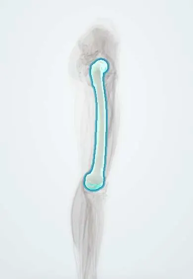
Fat embolism syndrome happens when fat from broken long bones enters the blood and lodges in the lungs, brain and skin, usually one to two days after injury.
Updated 2026-11-01
Structures involved
Tap one to see what it is and open it in 3D.
Why it happens
- Fat, air and amniotic fluid embolism
- The causes of hypoxemia
- Ventilation–perfusion matching and West zones
What it is
A clinical syndrome of respiratory distress, neurological changes and a petechial rash caused by fat globules entering the circulation, typically 24–72 hours after a long-bone or pelvic fracture. Fat embolism itself is common after these fractures; the full syndrome is much less frequent.
Who gets it
Reported incidence ranges from under 1% to more than 10% of patients with long-bone fractures, depending on the criteria and study design. It mainly affects young adults with femoral or tibial shaft fractures, a common pattern after motorcycle crashes in Colombia and Latin America.
Causes
- Closed fractures of the femoral or tibial shaft
- Pelvic fractures and multiple fractures
- Intramedullary reaming and nailing, and joint replacement
- Less often: liposuction, burns, bone marrow harvest, pancreatitis and sickle cell crisis with bone marrow infarction
Risk factors
- Young adult male
- Multiple long-bone fractures
- Closed rather than open fractures
- Delayed fracture stabilisation or prolonged traction
- Hypovolaemic shock and polytrauma
Pathophysiology
In the mechanical theory, raised pressure in the marrow cavity pushes fat droplets into torn venous sinusoids; they lodge in pulmonary capillaries and can pass to the brain and skin through the lungs or a patent foramen ovale. In the biochemical theory, the fat is broken down into free fatty acids that damage the capillary endothelium and trigger inflammation, causing lung injury that can progress to ARDS.
Classification
- Gurd and Wilson criteria: major (respiratory insufficiency, cerebral involvement, petechial rash) and minor (fever, tachycardia, retinal changes, jaundice, renal changes, anaemia, thrombocytopenia, raised ESR, fat macroglobulinaemia); diagnosis needs 2 major, or 1 major plus 4 minor
- Schonfeld and Lindeque scoring systems
- Subclinical, classic and fulminant forms
Symptoms and signs
- Shortness of breath, tachypnoea and low oxygen saturation
- Confusion, agitation, drowsiness or focal deficits
- Petechial rash on the upper chest, neck, axillae and conjunctivae
- Fever and tachycardia
- Retinal haemorrhages and fat globules on fundoscopy
Diagnosis
- Clinical
- A clinical diagnosis of exclusion in a patient with a recent long-bone or pelvic fracture, supported by the classic triad of hypoxaemia, neurological changes and petechiae and by criteria such as Gurd and Wilson.
- Tests
- Arterial blood gas (hypoxaemia), full blood count (anaemia, thrombocytopenia), coagulation studies, and sometimes fat globules in urine or bronchoalveolar lavage; none is specific.
- Imaging
- Chest X-ray may be normal or show bilateral patchy infiltrates. CT of the chest shows ground-glass opacities; CT pulmonary angiography helps exclude a thrombotic pulmonary embolism. Brain MRI shows multiple small diffusion-restricting lesions (starfield pattern) in cerebral fat embolism.
Differential diagnosis
- Pulmonary thromboembolism
- ARDS from other causes such as sepsis, aspiration or transfusion
- Lung contusion
- Traumatic brain injury
- Disseminated intravascular coagulation
Goal of treatment
Support oxygenation and organ function until the condition resolves, and lower risk through early fracture stabilisation.
Treatment
- Acute
- Supportive care: oxygen, non-invasive or mechanical ventilation with lung-protective settings and PEEP, careful fluid management, and haemodynamic support in the intensive care unit.
- Surgical
- Early stabilisation of long-bone fractures (within about 24 hours when the patient allows) reduces the risk; reaming techniques that limit marrow pressure are used during nailing.
- Medical
- No specific drug therapy has proven benefit; prophylactic corticosteroids remain controversial, and anticoagulants are not used as a treatment for the fat emboli.
- Follow-up
- Nursing priorities in fracture patients during the first 72 hours: pulse oximetry, respiratory rate, level of consciousness and a skin check for petechiae, with immobilisation of the fracture and early reporting of changes.
Complications
- Acute respiratory distress syndrome
- Cerebral oedema, seizures and coma
- Right heart failure
- Disseminated intravascular coagulation
- Death in fulminant cases
Prognosis
Most patients recover fully with supportive care, including neurological recovery; reported mortality with modern intensive care is under 10%.
Prevention
- Early immobilisation and early surgical fixation of long-bone fractures
- Careful operative technique to limit intramedullary pressure
- Close monitoring of high-risk fracture patients
Test yourself
Think of the answer, then tap the question to see it.
What is the classic triad of fat embolism syndrome?
Respiratory distress (hypoxaemia), neurological changes and a petechial rash.
When does fat embolism syndrome typically appear after a femur fracture?
Usually 24–72 hours after the injury.
Where is the petechial rash of fat embolism syndrome typically found?
Upper chest, neck, axillae and conjunctivae.
Which surgical measure lowers the risk of fat embolism syndrome?
Early stabilisation (fixation) of long-bone fractures.
What brain MRI pattern suggests cerebral fat embolism?
Multiple small diffusion-restricting lesions scattered through the brain, called the starfield pattern.
Related conditions
- Femoral shaft fracture
- Tibial shaft fracture
- Major trauma (polytrauma): ATLS primary survey
- Acute respiratory distress syndrome (ARDS)
- Pulmonary embolism
- Deep vein thrombosis
- Traumatic brain injury
- Sickle cell disease
Educational information, not medical advice. For any symptom, see a health professional.
Sources: Fat Embolism Syndrome – A Qualitative Review of its Incidence, Presentation, Pathogenesis and Management (Malaysian Orthopaedic Journal, 2021).
Spotted a mistake?We are always improving for the community. Tell us what is wrong and our team will review it.Report a mistake
Representative model, for educational purposes only.