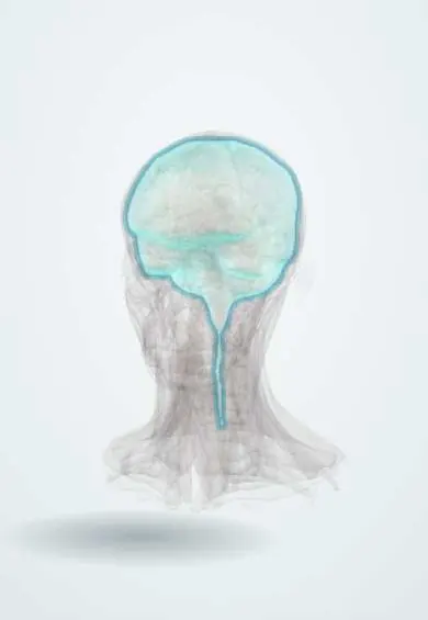
Traumatic brain injury !
A traumatic brain injury is brain damage from a blow, fall or jolt to the head; it ranges from a mild concussion to bleeding and coma.
Updated 2026-10-29
Structures involved
Tap one to see what it is and open it in 3D.
- Dura mater
- Frontal lobe
- Temporal lobe
- Middle meningeal artery
- Temporal bones
- Pterion
- Superior sagittal sinus
- Corpus callosum
- Oculomotor nerve (III)
- Midbrain
In the clinical exam
- Level of consciousness and the Glasgow Coma Scale
- The neurological examination: order and screening exam
- Pupils: light reflex and abnormal pupils (Marcus Gunn, Horner, Argyll Robertson)
- Intracranial hypertension syndrome
In physiology
- Nervous control of the circulation and the CNS ischemic response
- Cerebral blood flow, brain metabolism and intracranial pressure
- Wakefulness, sleep, the EEG and seizures
- Learning and memory
- Taste and smell
Why it happens
- Mechanical and electrical injury: abrasion, contusion, laceration, wounds
- Gray and white matter, cerebellar cortex, meninges
What it is
Brain damage caused by an external force such as a blow, fall or acceleration–deceleration. It ranges from concussion to severe injury with haematomas and coma.
Who gets it
A major cause of death and disability in young people. In Colombia and Latin America, road traffic crashes, especially those involving motorcycles, falls and violence are important causes.
Causes
- Road traffic crashes, especially motorcyclists and pedestrians
- Falls (children and older adults)
- Assaults and gunshot wounds
- Sports injuries
- Abusive head trauma in infants
Risk factors
- Not wearing a helmet or seat belt
- Alcohol and drug use
- Young male sex
- Older age and anticoagulant use (bleeding risk)
- Previous head injury
Pathophysiology
Primary injury occurs at impact: contusions, haematomas and diffuse axonal shearing. Secondary injury follows over hours to days from hypoxia, low blood pressure, swelling and raised intracranial pressure, which reduce brain perfusion. Preventing secondary injury is the main target of care.
Classification
- By Glasgow Coma Scale: mild 13–15, moderate 9–12, severe 3–8
- Epidural haematoma: arterial, often middle meningeal artery after temporal fracture; lens-shaped, does not cross sutures
- Subdural haematoma: torn bridging veins; crescent-shaped, crosses sutures; common in older adults
- Contusion, traumatic SAH and diffuse axonal injury
- Concussion: transient brain dysfunction without structural lesion on imaging
Symptoms and signs
- Headache, confusion, amnesia for the event
- Loss of consciousness
- Vomiting
- Dizziness, poor concentration, sleep problems (concussion)
- Focal deficits, seizures
- Lucid interval then deterioration (classically epidural haematoma)
Diagnosis
- Clinical
- Trauma primary survey (airway with cervical spine protection, breathing, circulation), then Glasgow Coma Scale and pupils, repeated over time. Clinical decision rules guide CT use in minor head injury.
- Tests
- Glucose, blood count, clotting, blood alcohol and toxicology when relevant; blood gas in severe injury.
- Imaging
- Non-contrast head CT is the first study for haematomas, fractures and swelling; cervical spine imaging as indicated. MRI shows diffuse axonal injury better.
Differential diagnosis
- Intoxication (alcohol, drugs)
- Hypoglycaemia
- Stroke or seizure that caused the fall
- Spontaneous intracranial haemorrhage
Goal of treatment
Prevent secondary brain injury by keeping oxygen, blood pressure and brain perfusion normal; remove mass lesions early; and support recovery.
Treatment
- Acute
- ATLS approach: secure the airway (intubation if GCS is 8 or less), avoid hypoxia and hypotension, protect the cervical spine.
- Surgical
- Craniotomy to evacuate significant epidural or subdural haematomas; decompressive craniectomy for refractory raised pressure in selected cases.
- Follow-up
- Intensive care: intracranial pressure monitoring in selected severe cases, head elevation, osmotic therapy, normal temperature, glucose and sodium.
- Medical
- Short-term antiseizure medication in severe injury; reversal of anticoagulants; analgesia.
- Conservative
- Concussion: brief relative rest followed by graded return to activity and study or sport.
- Rehabilitation
- Physical, cognitive and speech rehabilitation.
Drugs for this condition
Complications
- Brain herniation
- Post-traumatic epilepsy
- Chronic subdural haematoma
- Post-concussion syndrome
- Cognitive, behavioural and mood changes
- CSF leak and meningitis after skull base fracture
Prognosis
Most mild injuries recover over days to weeks, though some have lasting symptoms. In severe injury, outcome depends on age, initial Glasgow Coma Scale, pupils, CT findings and avoidance of hypoxia and hypotension.
Prevention
- Helmet use on motorcycles and bicycles
- Seat belts, child seats and road safety measures
- Fall prevention in older adults
- Not driving after alcohol
Test yourself
Think of the answer, then tap the question to see it.
What Glasgow Coma Scale score defines severe traumatic brain injury?
A score of 8 or less.
Which artery is typically torn in an epidural haematoma?
The middle meningeal artery, often with a temporal bone fracture near the pterion.
How do epidural and subdural haematomas look on CT?
Epidural: lens-shaped (biconvex), does not cross sutures. Subdural: crescent-shaped, crosses sutures.
What is the Cushing triad?
Hypertension, bradycardia and irregular breathing, a late sign of raised intracranial pressure.
Which two physiological insults most worsen outcome after brain injury?
Hypoxia and hypotension.
Related conditions
- Intracerebral haemorrhage
- Subarachnoid haemorrhage
- Epilepsy and seizures
- Spinal cord injury
- Epidural haematoma
- Subdural haematoma
- Mandible fracture
- Orbital floor (blowout) fracture
Educational information, not medical advice. For any symptom, see a health professional.
Sources: Traumatic Brain Injury (StatPearls (NCBI Bookshelf), 2025).
Spotted a mistake?We are always improving for the community. Tell us what is wrong and our team will review it.Report a mistake
Representative model, for educational purposes only.