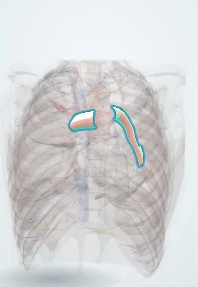
Pulmonary arteries !
The pulmonary arteries are the right and left branches of the pulmonary trunk; they deliver oxygen-poor blood into each lung to pick up oxygen.
Official name: Right pulmonary artery
Opens in the Durgical app, no sign-up needed.
What it is
The right and left branches of the pulmonary trunk, one to each lung, dividing to follow the bronchi.
What it does
Deliver oxygen-poor blood to the lungs to pick up oxygen.
Key facts
- Origin
- Bifurcation of pulmonary trunk; right passes behind ascending aorta and SVC; left joined to arch by ligamentum arteriosum
- Branches
- Lobar then segmental arteries, running with the bronchi
- Supplies
- Alveolar capillaries for gas exchange (lung tissue itself fed by bronchial arteries)
Relations
- Course
- Right: under the aortic arch, behind the ascending aorta and superior vena cava, in front of the right main bronchus, giving its upper lobe branch before the hilum; left: up and back over the left main bronchus, in front of the descending thoracic aorta
- In front
- Right: ascending aorta, superior vena cava and the right superior pulmonary vein; left: the left superior pulmonary vein
- Behind
- Right: right main bronchus and bronchus intermedius; left: left main bronchus and descending thoracic aorta
- Above
- Right: the arch of the azygos vein above the hilum; left: the aortic arch, tied to it by the ligamentum arteriosum, with the left recurrent laryngeal nerve hooking under the arch
- Below
- Right: the roof of the left atrium, with the transverse pericardial sinus just below and in front; left: the left main bronchus, over which it arches
How to find it
Not palpable. In a dissection of the hilum the artery is the highest structure on the left and lies in front of the bronchus on the right, with the pulmonary veins lowest and most anterior. On CT the arteries are traced alongside the bronchi.
On imaging
CT pulmonary angiography shows emboli as filling defects. On a frontal chest X-ray a widened right descending (interlobar) pulmonary artery suggests pulmonary hypertension; with embolism there may be an abrupt cut-off with a clear, under-perfused area beyond it (Westermark sign) or a peripheral wedge (Hampton hump).
Common problems
- Pulmonary embolism: a clot, usually from a leg vein, lodges here; causes sudden breathlessness and chest pain and can be fatal.
- Pulmonary hypertension: high pressure that strains the right ventricle.
How it is diagnosed
CT pulmonary angiography; D-dimer blood test; echocardiogram.
Treatment
- Blood thinners; clot-dissolving drugs or catheter removal for large clots.
Clinical notes
- The lower lobes receive most of the blood flow, so emboli lodge more often in lower lobe arteries
- A balloon-tipped pulmonary artery catheter passes through the right heart into a branch; its wedge pressure estimates left atrial pressure
- Mediastinal tumours or nodes can compress a pulmonary artery, reducing flow to one lung
Anatomical variants
Development
They come from the 6th pharyngeal arch arteries. The left 6th arch also forms the ductus arteriosus, which is why the left recurrent laryngeal nerve hooks under the ligamentum arteriosum and the aortic arch.
Memory aid
RALS: Right Anterior, Left Superior, the position of the pulmonary artery relative to the main bronchus at each hilum.
Related conditions
- Pulmonary hypertension and cor pulmonale
- Eisenmenger syndrome
- High-altitude illness (AMS, HACE, HAPE)
- Persistent pulmonary hypertension of the newborn
- Pulmonary embolism
Test yourself
Think of the answer, then tap the question to see it.
At the hilum, where does the pulmonary artery lie relative to the main bronchus on each side?
In front of the bronchus on the right and above it on the left; the pulmonary veins are the lowest and most anterior.
How do the right and left pulmonary arteries differ in course?
The right is longer and runs horizontally under the aortic arch behind the ascending aorta and superior vena cava; the left is shorter and arches over the left main bronchus.
What are the Westermark sign and Hampton hump on a chest X-ray?
Signs of pulmonary embolism: an abrupt arterial cut-off with a clear, under-perfused area beyond it (Westermark), and a peripheral wedge-shaped opacity (Hampton hump).
Why do pulmonary emboli lodge more often in the lower lobe arteries?
Because the lower lobes receive most of the pulmonary blood flow.
From which pharyngeal arch arteries do the pulmonary arteries develop?
The 6th; the left 6th also forms the ductus arteriosus, so the left recurrent laryngeal nerve hooks under the ligamentum arteriosum.
Educational information, not medical advice. For any symptom, see a health professional.
Its names
- Official Latin (TA2)
- arteria pulmonalis dextra; arteria pulmonalis sinistra TA2 4077
- Official Spanish (DeCS)
- Arteria pulmonar DeCS D011651
- English (TA2)
- Right pulmonary artery
- Also called
- ramus dexter arteriae pulmonalis
Sources: Terminologia Anatomica (TA2); DeCS; Anatomy, Thorax, Heart Great Vessels; Anatomy, Thorax, Lungs; Tratado de anatomía humana (9.ª ed.), tomo II, Libro IV, sección 2 Arterias, caps. I-III (sistema de la arteria pulmonar; aorta y ramas del cayado hasta la carótida interna).
Spotted a mistake?We are always improving for the community. Tell us what is wrong and our team will review it.Report a mistake
Representative model, for educational purposes only.