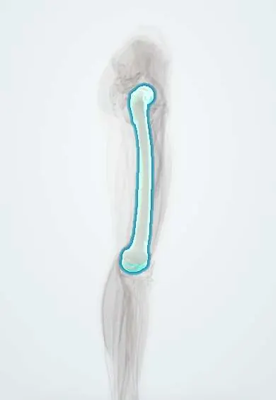
Femoral shaft fracture !
A femoral shaft fracture is a break in the long middle part of the thigh bone, usually from high-energy trauma such as a traffic crash.
Updated 2026-10-29
Structures involved
Tap one to see what it is and open it in 3D.
- Femur
- Femoral artery
- Deep femoral artery
- Sciatic nerve
- Vastus intermedius
- Vastus lateralis
- Adductor magnus
Why it happens
What it is
A fracture of the diaphysis of the femur, between the region below the lesser trochanter and the area just above the condyles, usually caused by high-energy trauma.
Who gets it
Mostly young adults after traffic collisions (including motorcycle crashes, very common in Colombia), falls from height or gunshot wounds; in older people a low-energy fall on weak bone can be enough.
Causes
- High-energy trauma: traffic collisions, motorcycle crashes, falls from height
- Gunshot wounds
- Low-energy falls in osteoporotic bone
- Pathological fracture through a tumour or metastasis
- Atypical femoral fracture after long-term antiresorptive therapy
Risk factors
- Motorcycle use and road traffic exposure
- Osteoporosis
- Bone metastases
- Long-term bisphosphonate therapy (atypical fractures)
- Non-accidental injury in young children
Pathophysiology
The femoral shaft is the strongest bone in the body, so breaking it takes a lot of force and associated injuries are common. The thigh can hide a large amount of blood, enough to cause shock. Marrow fat released into the circulation can lodge in the lungs, brain and skin (fat embolism).
Classification
- By location: proximal, middle or distal third
- By pattern: transverse, oblique, spiral, comminuted or segmental
- Winquist-Hansen grading of comminution
- Open or closed (Gustilo-Anderson for open fractures)
Symptoms and signs
- Severe thigh pain after trauma
- Swelling and deformity of the thigh
- Shortening and rotation of the limb
- Inability to stand or move the leg
Diagnosis
- Clinical
- Trauma assessment by the ATLS approach first, then examination of the limb, distal pulses and nerves (sciatic, tibial and fibular), and the skin for open wounds.
- Tests
- Blood count, cross-match and lactate in trauma; arterial blood gas if fat embolism is suspected.
- Imaging
- Radiographs of the entire femur plus the hip and knee; CT of the femoral neck to exclude an associated fracture; angiography or CT angiography if a vascular injury is suspected.
Differential diagnosis
- Femoral neck or intertrochanteric fracture
- Distal femur (supracondylar) fracture
- Hip dislocation
- Pathological fracture
- Thigh muscle injury or haematoma
Goal of treatment
Resuscitate the patient, stabilise the fracture early to reduce blood loss and pulmonary complications, restore length, alignment and rotation, and get the patient moving.
Treatment
- Acute
- Primary survey and resuscitation with fluids and blood products; analgesia, including a femoral nerve or fascia iliaca block.
- Acute
- Temporary traction splint or skeletal traction to reduce pain and bleeding until surgery.
- Surgical
- Intramedullary nailing within the first day or so in stable patients; the standard treatment in adults.
- Surgical
- External fixation as damage control in unstable polytrauma, converted to a nail later.
- Medical
- Antibiotics and debridement for open fractures; thromboprophylaxis with anticoagulants.
- Conservative
- Spica cast or flexible nails in young children, depending on age and weight.
- Rehabilitation
- Early weight-bearing as tolerated and physiotherapy for knee and hip motion.
Complications
- Haemorrhagic shock
- Fat embolism syndrome
- Deep vein thrombosis and pulmonary embolism
- Associated femoral neck fracture missed at first
- Compartment syndrome
- Infection, delayed union, nonunion or malunion (rotation or length)
- Knee stiffness
Prognosis
With intramedullary nailing most fractures unite, and most patients regain walking. Outcomes depend on associated injuries, open wounds and age.
Prevention
- Road safety measures: helmets, speed limits and seat belts
- Osteoporosis treatment and fall prevention in older adults
Test yourself
Think of the answer, then tap the question to see it.
What is the standard treatment for a femoral shaft fracture in an adult?
Early intramedullary nailing, once the patient is stable.
Which complication causes confusion, hypoxaemia and petechiae 1 to 3 days after a femoral shaft fracture?
Fat embolism syndrome, from marrow fat entering the circulation.
Why can a closed femoral shaft fracture cause shock?
The thigh can hold a large volume of blood, enough to cause hypovolaemic shock.
Which associated fracture must be excluded in every femoral shaft fracture?
An ipsilateral femoral neck fracture, which is easy to miss.
What does early fracture stabilisation reduce in femoral shaft fractures?
Pulmonary complications such as fat embolism and ARDS, plus pain, bleeding and immobility.
Related conditions
- Hypovolaemic shock
- Acute compartment syndrome
- Deep vein thrombosis
- Hip dislocation
- Femoral Neck Fracture
- Pulmonary embolism
Educational information, not medical advice. For any symptom, see a health professional.
Sources: Femoral Shaft Fractures (StatPearls (NCBI Bookshelf), 2023).
Spotted a mistake?We are always improving for the community. Tell us what is wrong and our team will review it.Report a mistake
Representative model, for educational purposes only.