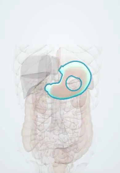
Helicobacter pylori gastritis !
Helicobacter pylori gastritis is long-term stomach inflammation caused by a common bacterium; it causes most peptic ulcers and raises stomach cancer risk.
Updated 2026-10-29
Structures involved
Tap one to see what it is and open it in 3D.
In biochemistry
Microorganisms that cause it
In histology
In physiology
Why it happens
- Chronic inflammation: causes, cells and features
- Metaplasia
- Multistep carcinogenesis: the adenoma–carcinoma sequence
- Diet, cancer and systemic disease
- Precancerous conditions: when chronic inflammation, metaplasia or hyperplasia raise the risk
- Oncogenic viruses and H. pylori: the table to know
What it is
Chronic inflammation of the stomach lining caused by Helicobacter pylori, a spiral, urease-producing bacterium. It causes most peptic ulcers and is a major cause of gastric cancer.
Who gets it
One of the most common chronic infections in the world, usually acquired in childhood. It is very common in Colombia and the rest of Latin America, and the country's Andean highlands have some of its highest gastric cancer rates.
Causes
- Infection with Helicobacter pylori (gram-negative, flagellated, microaerophilic)
- Person-to-person spread (oral-oral, faecal-oral), mostly within families in childhood
- Urease, flagella and adhesins let it survive and attach in the acidic stomach
- Virulent strains (CagA, VacA) cause more inflammation
Risk factors
- Childhood in crowded housing or with limited sanitation
- Infected family members
- Lower socioeconomic conditions
- Living in regions with high prevalence
Pathophysiology
The bacterium lives beneath the mucus layer and triggers chronic active gastritis. Antrum-predominant gastritis raises acid output and leads to duodenal ulcers. Body-predominant gastritis lowers acid and can progress through atrophy and intestinal metaplasia to gastric adenocarcinoma (Correa cascade). It also drives gastric MALT lymphoma.
Classification
- Antrum-predominant versus corpus-predominant (pangastritis)
- Non-atrophic versus atrophic gastritis; staging systems such as OLGA
- Sydney system for histology
Symptoms and signs
- Most infected people have no symptoms
- Dyspepsia: epigastric pain or burning, fullness, early satiety, nausea
- Symptoms of peptic ulcer when one develops
- Unexplained iron deficiency anaemia or vitamin B12 deficiency
- Immune thrombocytopenia (association)
Diagnosis
- Clinical
- Testing is done when there is a reason to treat: peptic ulcer, MALT lymphoma, dyspepsia, family history of gastric cancer, long-term NSAID or aspirin use, unexplained iron deficiency or immune thrombocytopenia.
- Tests
- Non-invasive: urea breath test and stool antigen test. At endoscopy: biopsies for histology, rapid urease test, culture with antibiotic susceptibility. Serology cannot tell current from past infection. Proton pump inhibitors, antibiotics and bismuth cause false negatives and are stopped beforehand.
- Imaging
- Imaging is not used to detect the infection. Endoscopy with biopsies is preferred when there are alarm features or higher cancer risk.
Differential diagnosis
- Functional dyspepsia
- NSAID-induced gastropathy
- Gastro-oesophageal reflux disease
- Gastric cancer
- Biliary colic
- Autoimmune atrophic gastritis
Goal of treatment
Eradicate the bacterium, heal ulcers, prevent recurrence and bleeding and lower the risk of gastric cancer, then confirm that eradication worked.
Treatment
- Medical
- Eradication therapy: a proton pump inhibitor combined with two or three antibiotics (penicillin, macrolide, nitroimidazole, tetracycline classes), with or without bismuth, usually for 14 days.
- Medical
- The regimen is chosen by local antibiotic resistance; where macrolide resistance is high, bismuth-based quadruple therapy is generally preferred. Guidelines vary.
- Medical
- Second-line or culture-guided therapy when the first treatment fails.
- Follow-up
- Confirm eradication with a urea breath test or stool antigen at least four weeks after treatment, off acid suppression.
- Follow-up
- Endoscopic surveillance for extensive atrophy or intestinal metaplasia.
Drugs for this condition
Complications
- Duodenal and gastric ulcers
- Upper gastrointestinal bleeding and perforation
- Atrophic gastritis and intestinal metaplasia
- Gastric adenocarcinoma
- Gastric MALT lymphoma
- Iron and vitamin B12 deficiency
Prognosis
Eradication heals most ulcers and markedly lowers ulcer recurrence; early-stage MALT lymphoma often regresses. Eradication before atrophy develops lowers gastric cancer risk the most. Reinfection is possible where the infection is common.
Prevention
- Safe drinking water, sanitation and hygiene
- Test-and-treat strategies in high-risk groups
- Screening of family members of gastric cancer patients in some guidelines
Test yourself
Think of the answer, then tap the question to see it.
Which non-invasive tests detect active H. pylori infection?
The urea breath test and the stool antigen test. Serology only shows past or present exposure.
Why are proton pump inhibitors stopped before testing for H. pylori?
They suppress the bacterium and can give false-negative breath, stool and urease tests.
What sequence links H. pylori to intestinal-type gastric cancer?
Correa cascade: chronic gastritis, atrophic gastritis, intestinal metaplasia, dysplasia, adenocarcinoma.
Which lymphoma can regress after H. pylori eradication alone?
Low-grade gastric MALT lymphoma, when it is localised and H. pylori positive.
What determines the choice of eradication regimen?
Local antibiotic resistance (especially to macrolides), previous antibiotic use and patient allergy; bismuth quadruple therapy is common where resistance is high.
Related conditions
- Peptic ulcer disease
- Gastric cancer
- Upper gastrointestinal bleeding
- Iron deficiency anaemia
- Gastro-oesophageal reflux disease (GERD)
Educational information, not medical advice. For any symptom, see a health professional.
Sources: Helicobacter Pylori (StatPearls (NCBI Bookshelf), 2026).
Spotted a mistake?We are always improving for the community. Tell us what is wrong and our team will review it.Report a mistake
Representative model, for educational purposes only.