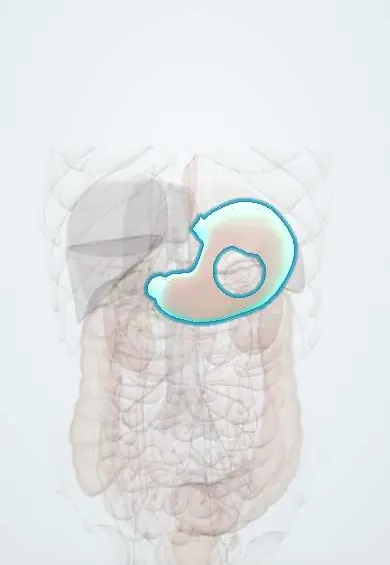
Gastric cancer !
Gastric cancer is a malignant tumour of the stomach, linked to Helicobacter pylori; it often causes vague symptoms and is found late. It is common in Colombia.
Updated 2026-10-29
Structures involved
Tap one to see what it is and open it in 3D.
- Stomach
- Celiac and gastric lymph nodes
- Gastric arteries
- Lesser omentum
- Greater omentum
- Esophagus
- Duodenum
- Liver
- Peritoneum
In biochemistry
Microorganisms that cause it
In histology
- Cell junctions: tight, adherens, desmosomes, gap, hemidesmosomes
- Stomach: gastric glands, parietal and chief cells
In the clinical exam
Why it happens
- Metaplasia
- Precancerous conditions: when chronic inflammation, metaplasia or hyperplasia raise the risk
- Cancer epidemiology: age, environment, predisposing conditions (and Colombia)
- Diet, cancer and systemic disease
- Local effects of tumours and cancer cachexia
- Multistep carcinogenesis: the adenoma–carcinoma sequence
Show 7 more
- Pathways of spread and the usual metastatic sites
- Grading and staging: what TNM means
- From specimen to report: fixation, margins and the pathology report
- Invasion and metastasis: the metastatic cascade, step by step
- Chemical carcinogens: initiation, promotion and the table to know
- Chronic inflammation: causes, cells and features
- Oncogenic viruses and H. pylori: the table to know
What it is
A malignant tumour of the stomach, almost always an adenocarcinoma, that often causes vague symptoms and is diagnosed late.
Who gets it
Among the most frequent causes of cancer death worldwide. Latin America, including Colombia, has some of the highest rates, with particularly high incidence reported in Andean mountain populations, linked to H. pylori infection and diet. It is more common in men and after middle age.
Causes
- Chronic Helicobacter pylori infection (the main cause)
- Correa cascade: chronic gastritis, atrophy, intestinal metaplasia, dysplasia and cancer
- Hereditary diffuse gastric cancer (CDH1 mutation), Lynch syndrome and other inherited syndromes
- Epstein-Barr virus-associated tumours
Risk factors
- H. pylori infection, especially acquired in childhood
- Diet high in salt and in salted, smoked or cured foods; low fruit and vegetable intake
- Smoking and heavy alcohol use
- First-degree relative with gastric cancer
- Atrophic gastritis (including autoimmune / pernicious anaemia), intestinal metaplasia, adenomatous polyps
- Previous partial gastrectomy; male sex and older age
Pathophysiology
Long-standing H. pylori inflammation drives a stepwise sequence from atrophic gastritis to intestinal metaplasia, dysplasia and the intestinal type of adenocarcinoma. The diffuse type (signet-ring cells) arises without this sequence, often from loss of E-cadherin, and spreads through the wall (linitis plastica). Spread is by lymphatics, the peritoneum and the portal vein to the liver.
Classification
- Lauren: intestinal vs diffuse (and mixed)
- Early gastric cancer (limited to mucosa or submucosa) vs advanced
- Borrmann macroscopic types I-IV for advanced tumours
- TNM staging; Siewert classification for tumours at the gastro-oesophageal junction
Symptoms and signs
- Often no symptoms in early stages
- Persistent dyspepsia or epigastric pain
- Early satiety, loss of appetite and unintended weight loss
- Nausea and vomiting (gastric outlet obstruction with distal tumours)
- Difficulty swallowing (proximal tumours)
- Iron deficiency anaemia, melaena or haematemesis
- Signs of spread: left supraclavicular node (Virchow), periumbilical nodule (Sister Mary Joseph), ovarian metastasis (Krukenberg), ascites
Diagnosis
- Clinical
- Alarm features in a patient with dyspepsia, especially from high-incidence areas or with family history. Examination for nodes, masses, ascites and liver enlargement.
- Tests
- Upper endoscopy with multiple biopsies is the diagnostic test; histology gives the type and HER2 and mismatch repair or PD-L1 status guide therapy. Blood count, iron studies, liver tests; H. pylori testing.
- Imaging
- CT of chest, abdomen and pelvis for staging. Endoscopic ultrasound assesses depth and nearby nodes; PET-CT in selected cases. Staging laparoscopy (with peritoneal washings) looks for peritoneal spread before major surgery.
Differential diagnosis
- Peptic ulcer disease
- Functional dyspepsia and chronic gastritis
- Gastric lymphoma (MALT) and gastrointestinal stromal tumour
- Oesophageal cancer
- Pancreatic or biliary cancer
Goal of treatment
Cure localised disease by complete resection with adequate lymph node removal; in advanced disease, prolong survival and relieve bleeding, obstruction and pain.
Treatment
- Procedure
- Endoscopic resection (endoscopic mucosal resection or submucosal dissection) for selected early cancers confined to the mucosa with low node risk.
- Surgical
- Surgical resection: subtotal (distal) or total gastrectomy with D2 lymphadenectomy, open or minimally invasive, for resectable tumours.
- Medical
- Perioperative or adjuvant chemotherapy for locally advanced cancer; regimens and use of chemoradiotherapy vary between guidelines.
- Medical
- Advanced disease: systemic chemotherapy combined, according to the tumour profile, with HER2-targeted antibodies or immune checkpoint inhibitors.
- Procedure
- Palliation: endoscopic stents or surgical bypass for obstruction, radiotherapy or endoscopy for bleeding.
- Medical
- Eradication of H. pylori after resection of early cancer and in first-degree relatives, plus nutritional support and vitamin B12 replacement after gastrectomy.
Drugs for this condition
Complications
- Gastric outlet obstruction
- Bleeding and iron deficiency anaemia
- Perforation
- Peritoneal carcinomatosis and malignant ascites
- Liver metastases
- After gastrectomy: dumping syndrome, vitamin B12 and iron deficiency, weight loss, anastomotic leak
Prognosis
Early gastric cancer has a very good outlook after resection. Most cases are found at an advanced stage, when survival is poor; stage is the main prognostic factor.
Prevention
- Treatment of H. pylori infection, which reduces gastric cancer risk
- Diet lower in salt and preserved foods, with fresh fruit and vegetables; not smoking
- Endoscopic surveillance of extensive atrophy or intestinal metaplasia; screening programmes in high-risk areas vary
Test yourself
Think of the answer, then tap the question to see it.
Which infection is the main risk factor for gastric adenocarcinoma?
Chronic Helicobacter pylori infection.
What sequence leads to the intestinal type of gastric cancer (Correa cascade)?
Chronic gastritis, atrophic gastritis, intestinal metaplasia, dysplasia and then adenocarcinoma.
What are the two Lauren histological types of gastric adenocarcinoma?
Intestinal and diffuse; the diffuse type has signet-ring cells and can cause linitis plastica.
What is the Virchow node and why is it relevant in gastric cancer?
An enlarged left supraclavicular lymph node, a sign of metastatic spread of abdominal cancer.
Which test confirms the diagnosis of gastric cancer?
Upper endoscopy with biopsies of the lesion.
Related conditions
- Peptic ulcer disease
- Upper gastrointestinal bleeding
- Helicobacter pylori gastritis
- Oesophageal cancer
- Ovarian cancer
Educational information, not medical advice. For any symptom, see a health professional.
Sources: Gastric Cancer (StatPearls (NCBI Bookshelf), 2024).
Spotted a mistake?We are always improving for the community. Tell us what is wrong and our team will review it.Report a mistake
Representative model, for educational purposes only.