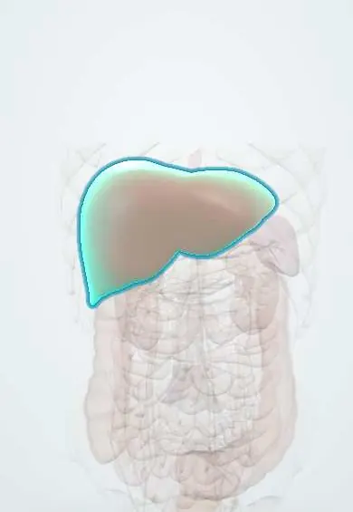
Hepatocellular carcinoma !
Hepatocellular carcinoma is the main primary liver cancer, arising from liver cells, usually in a liver with cirrhosis or chronic hepatitis B.
Updated 2026-10-29
Structures involved
Tap one to see what it is and open it in 3D.
- Liver
- Portal vein
- Hepatic veins
- Proper hepatic artery
- Common hepatic artery
- Inferior vena cava
- Celiac and gastric lymph nodes
- Spleen
In biochemistry
- Liver function tests: reading the pattern
- Tumour markers: principles of use
- Alpha-fetoprotein (AFP)
- δ-Aminolevulinic acid (ALA)
- α1-antitrypsin
Microorganisms that cause it
In histology
In the clinical exam
Why it happens
- Precancerous conditions: when chronic inflammation, metaplasia or hyperplasia raise the risk
- Tumour angiogenesis
- Chemical carcinogens: initiation, promotion and the table to know
- Diet, cancer and systemic disease
- Paraneoplastic syndromes: the table to know
- Tumour markers: serum and tissue, and what each one is for
Show 6 more
- Liver regeneration
- Oncogenic viruses and H. pylori: the table to know
- Naming tumours: -oma, carcinoma, sarcoma, and the misleading names
- Cancer epidemiology: age, environment, predisposing conditions (and Colombia)
- Pathways of spread and the usual metastatic sites
- Alcohol: how it injures the liver, brain, heart and fetus
What it is
The main primary cancer of the liver, arising from hepatocytes, usually in a cirrhotic liver or in chronic hepatitis B.
Who gets it
A leading cause of cancer death worldwide. In Latin America it is linked mainly to cirrhosis from hepatitis B and C, alcohol and, increasingly, MASLD with obesity and diabetes; many patients are diagnosed late.
Causes
- Cirrhosis of any cause
- Chronic hepatitis B, even without cirrhosis
- Chronic hepatitis C
- Alcohol-related liver disease and MASLD
- Aflatoxin exposure, haemochromatosis, alpha-1 antitrypsin deficiency
Risk factors
- Cirrhosis (the strongest risk factor)
- Hepatitis B, especially with high viral load or family history of liver cancer
- Male sex and older age
- Obesity, type 2 diabetes, alcohol and smoking
Pathophysiology
Chronic inflammation and regeneration cause repeated cell turnover and genetic damage, producing dysplastic nodules that become cancer. The tumour takes its blood mostly from the hepatic artery, which explains arterial enhancement with washout on imaging. It tends to invade the portal and hepatic veins.
Classification
- BCLC (Barcelona Clinic Liver Cancer) staging: very early, early, intermediate, advanced and terminal
- LI-RADS categories (LR-1 to LR-5) for nodules in at-risk livers; LR-5 means definite HCC
- Milan criteria for transplantation (limited tumour size and number, no vascular invasion)
- Fibrolamellar carcinoma: a distinct variant in young people without cirrhosis
Symptoms and signs
- Often silent; found on surveillance
- Right upper quadrant pain or mass, weight loss, early satiety
- New decompensation of a stable cirrhosis (ascites, jaundice, encephalopathy, bleeding)
- Paraneoplastic features: hypoglycaemia, erythrocytosis, hypercalcaemia
Diagnosis
- Clinical
- Surveillance of at-risk patients, examination for hepatomegaly, ascites and signs of liver failure; liver reserve is graded with Child-Pugh and performance status.
- Tests
- Alpha-fetoprotein (AFP), raised in many but not all cases; liver tests, INR, albumin, hepatitis B and C serology. Biopsy if imaging is not typical or in a non-cirrhotic liver.
- Imaging
- Multiphase CT or MRI: arterial phase hyperenhancement with washout in later phases is diagnostic in a cirrhotic liver, reported with LI-RADS. Ultrasound is the surveillance test. Chest CT for staging.
Differential diagnosis
- Regenerative or dysplastic nodule
- Liver metastases
- Intrahepatic cholangiocarcinoma
- Haemangioma, focal nodular hyperplasia or adenoma
- Liver abscess
Goal of treatment
Cure the cancer when found early (resection, ablation or transplantation) and, at later stages, control the tumour and extend life while preserving liver function.
Treatment
- Surgical
- Liver resection for a single tumour in a patient with preserved liver function and no significant portal hypertension.
- Surgical
- Liver transplantation within accepted criteria (e.g., Milan), which treats both the cancer and the cirrhosis.
- Procedure
- Thermal ablation (radiofrequency or microwave) for small tumours.
- Procedure
- Transarterial chemoembolisation (TACE) or radioembolisation for intermediate-stage disease confined to the liver.
- Medical
- Advanced disease: immune checkpoint inhibitor combinations (often with an anti-VEGF agent) or tyrosine kinase inhibitors.
- Medical
- Antiviral treatment of hepatitis B or C and care of the underlying cirrhosis alongside cancer therapy.
Drugs for this condition
Complications
- Liver failure
- Portal vein tumour thrombosis and variceal bleeding
- Tumour rupture with haemoperitoneum
- Metastases to lung, bone and lymph nodes
- Recurrence after resection or ablation
Prognosis
Depends on tumour stage and liver function. Early tumours found by surveillance can be cured; most tumours found because of symptoms are advanced, with short survival.
Prevention
- Hepatitis B vaccination
- Treatment of hepatitis B and C
- Limiting alcohol; control of obesity and diabetes
- Ultrasound surveillance about every six months (with or without AFP) in cirrhosis and selected hepatitis B carriers
Test yourself
Think of the answer, then tap the question to see it.
Which imaging pattern is diagnostic of hepatocellular carcinoma in a cirrhotic liver?
Arterial phase hyperenhancement followed by washout in the portal venous or delayed phase on multiphase CT or MRI.
How are at-risk patients with cirrhosis screened for hepatocellular carcinoma?
Ultrasound about every six months, with or without serum alpha-fetoprotein.
Why can hepatitis B cause hepatocellular carcinoma without cirrhosis?
Its DNA can integrate into the host genome and promote cancer directly, besides causing chronic inflammation.
What does LR-5 mean in the LI-RADS system?
A nodule with imaging features of definite hepatocellular carcinoma, which can be treated without biopsy in an at-risk liver.
Which staging system links stage to treatment in hepatocellular carcinoma?
The BCLC (Barcelona Clinic Liver Cancer) system, which combines tumour burden, liver function and performance status.
Related conditions
Educational information, not medical advice. For any symptom, see a health professional.
Sources: Hepatocellular Carcinoma (StatPearls (NCBI Bookshelf), 2023).
Spotted a mistake?We are always improving for the community. Tell us what is wrong and our team will review it.Report a mistake
Representative model, for educational purposes only.