Inflammatory myopathies (dermatomyositis, polymyositis) !
Autoimmune diseases that inflame muscles and cause weakness of the shoulders and hips; dermatomyositis also has a typical rash.
Updated 2026-11-01
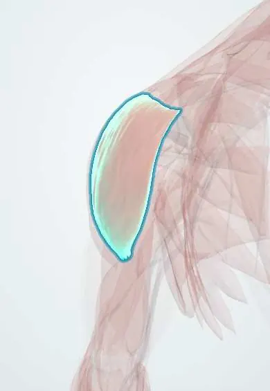
Structures involved
Tap one to see what it is and open it in 3D.
Why it happens
- Paraneoplastic syndromes: the table to know
- Creatine kinase (CK) and CK-MB as lab tests
- HMG-CoA reductase
- Skeletal muscle: sarcomere and contraction
- Muscle injury, repair and satellite cells
What it is
A group of autoimmune diseases (idiopathic inflammatory myopathies) that inflame skeletal muscle and cause symmetric proximal weakness. The main forms are dermatomyositis, polymyositis, immune-mediated necrotising myopathy and inclusion body myositis.
Who gets it
Rare. Dermatomyositis has two peaks, in children (juvenile dermatomyositis) and in adults around 40 to 60, and is more common in women. Inclusion body myositis is the most common inflammatory myopathy after age 50 and affects men more. Cases are seen in Colombia and the rest of Latin America in rheumatology, neurology and dermatology clinics.
Causes
- Autoimmune; cause unknown
- Underlying cancer in adult dermatomyositis (paraneoplastic)
- Statins can trigger anti-HMGCR necrotising myopathy
- Genetic factors and possibly viral infections or UV light as triggers
Risk factors
- Female sex (dermatomyositis, polymyositis)
- Male sex and age over 50 (inclusion body myositis)
- Cancer, especially ovarian, lung, gastric and nasopharyngeal, in adult dermatomyositis
- Other autoimmune diseases
Pathophysiology
In dermatomyositis, complement and antibodies damage small vessels around muscle fascicles, causing perifascicular atrophy, and the same vasculopathy affects skin. In polymyositis, cytotoxic CD8 T cells invade healthy muscle fibres. Necrotising myopathy shows muscle fibre death with few inflammatory cells. Inclusion body myositis combines T-cell inflammation with degenerative protein aggregates (rimmed vacuoles) and responds poorly to immunosuppression.
Classification
- Dermatomyositis: proximal weakness plus skin signs; amyopathic forms have the rash only
- Polymyositis: proximal weakness without rash; a diagnosis of exclusion
- Immune-mediated necrotising myopathy (anti-SRP, anti-HMGCR)
- Antisynthetase syndrome (anti-Jo-1): myositis, interstitial lung disease, arthritis, mechanic's hands, Raynaud phenomenon
- Inclusion body myositis: over 50, slow, asymmetric weakness of finger flexors and quadriceps
- Juvenile dermatomyositis: childhood form with frequent calcinosis and vasculopathy
Symptoms and signs
- Symmetric weakness of shoulders and hips over weeks to months: trouble climbing stairs, rising from a chair or lifting the arms
- Muscle aching or tenderness in some patients
- Dermatomyositis: heliotrope (violet) rash of the eyelids, Gottron papules over the knuckles, V-sign and shawl sign rash, nailfold changes
- Rough, cracked skin of the fingers (mechanic's hands)
- Difficulty swallowing (dysphagia)
- Breathlessness and cough from lung involvement
- Fever, fatigue and weight loss
Diagnosis
- Clinical
- Pattern of symmetric proximal weakness with or without characteristic skin lesions; manual muscle testing of neck flexors, deltoids and hip flexors. EULAR/ACR classification criteria combine clinical, laboratory and biopsy findings.
- Tests
- High CK, aldolase, AST, ALT and LDH. ANA and myositis-specific antibodies (anti-Jo-1, anti-Mi-2, anti-MDA5, anti-TIF1-gamma, anti-SRP, anti-HMGCR). Electromyography shows a myopathic pattern. Muscle biopsy is the reference test. Pulmonary function tests and ECG; age-appropriate cancer screening.
- Imaging
- MRI of the thigh muscles shows oedema and guides the biopsy site; high-resolution chest CT for interstitial lung disease; chest-abdomen-pelvis CT, mammography or pelvic ultrasound in the cancer search.
Differential diagnosis
- Hypothyroidism or other endocrine myopathy
- Drug or toxic myopathy (statins, glucocorticoids, alcohol)
- Muscular dystrophies
- Myasthenia gravis
- Polymyalgia rheumatica (pain without weakness)
- Electrolyte disorders (low potassium) and infections causing myositis
- Lupus or psoriasis for the skin findings
Goal of treatment
Recover strength and function, control skin and lung disease, protect swallowing and breathing, and find and treat any associated cancer.
Treatment
- Medical
- High-dose glucocorticoids are the initial treatment for dermatomyositis, polymyositis and necrotising myopathy, tapered as strength and CK improve.
- Medical
- Steroid-sparing immunosuppressants (methotrexate, azathioprine, mycophenolate); intravenous immunoglobulin, rituximab or calcineurin inhibitors for refractory, lung or swallowing involvement; hydroxychloroquine and sun protection for the rash.
- Rehabilitation
- Physiotherapy and an exercise programme to rebuild strength and prevent contractures; swallowing therapy.
- Follow-up
- Follow-up of strength, CK, lung function and swallowing; nursing care checks swallowing safety, aspiration risk, falls and skin integrity.
- Conservative
- Inclusion body myositis does not respond well to immunosuppressants; care is supportive with physiotherapy, mobility aids and swallowing support.
Complications
- Interstitial lung disease (especially antisynthetase and anti-MDA5 disease)
- Aspiration pneumonia from dysphagia
- Respiratory muscle failure
- Myocarditis and arrhythmias
- Associated cancer in adult dermatomyositis
- Calcinosis, especially in juvenile dermatomyositis
- Corticosteroid adverse effects and steroid myopathy
Prognosis
Most patients with dermatomyositis or polymyositis improve with treatment, though many keep some weakness and need long-term therapy. Cancer, lung disease and cardiac involvement worsen the outlook. Inclusion body myositis progresses slowly despite treatment.
Test yourself
Think of the answer, then tap the question to see it.
What skin findings are characteristic of dermatomyositis?
Heliotrope rash of the upper eyelids and Gottron papules over the knuckles; also V-sign, shawl sign and nailfold changes.
Why must adults with new dermatomyositis be screened for cancer?
Adult dermatomyositis is often paraneoplastic, notably with ovarian, lung, gastric and nasopharyngeal cancers.
What features define the antisynthetase syndrome?
Anti-Jo-1 (or other antisynthetase) antibodies with myositis, interstitial lung disease, arthritis, mechanic's hands and Raynaud phenomenon.
Which inflammatory myopathy affects older men, with finger flexor and quadriceps weakness and poor steroid response?
Inclusion body myositis.
What is the reference test to confirm an inflammatory myopathy?
Muscle biopsy, supported by CK, myositis antibodies, EMG and MRI.
Related conditions
- Interstitial lung disease
- Systemic sclerosis (scleroderma, including CREST)
- Systemic lupus erythematosus
- Polymyalgia rheumatica
- Hypothyroidism
- Lung cancer
- Ovarian cancer
Educational information, not medical advice. For any symptom, see a health professional.
Sources: Dermatomyositis (MedlinePlus (U.S. National Library of Medicine), 2025).
Spotted a mistake?We are always improving for the community. Tell us what is wrong and our team will review it.Report a mistake
Representative model, for educational purposes only.