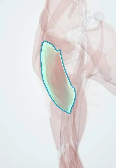
Rear deltoid !
The rear deltoid is the back part of the shoulder's rounded muscle; it pulls the arm back and turns it outward.
Official name: Deltoid, posterior (spinal) part
Opens in the Durgical app, no sign-up needed.
What it is
The back part of the deltoid; it pulls the arm back and turns it outward.
What it does
Pulls the arm back and turns it outward.
Key facts
- Origin
- Spine of the scapula
- Insertion
- Deltoid tuberosity of the humerus
- Action
- Extends and laterally rotates the arm
- Nerve
- Axillary nerve (C5–C6)
- Blood supply
- Posterior circumflex humeral artery, thoracoacromial artery (deltoid branch)
Relations
- Above
- Spine of the scapula
- Deep
- Infraspinatus, teres minor and the triceps (long head); the quadrangular space with the axillary nerve and posterior circumflex humeral vessels
- In front
- Middle deltoid
- Below
- Teres major and the triceps (lateral head); the upper lateral cutaneous nerve of the arm turns round its lower border
How to find it
With the arm out to the side at 90° and pulled backward against resistance, the posterior part tightens below the spine of the scapula, behind the shoulder.
Examination
- Deltoid extension lag sign: the examiner lifts the straight arm into near-full extension and lets go; if the arm drifts forward, the rear deltoid (axillary nerve) is weak
- Resisted horizontal extension at 90° abduction: weakness compared with the other side points to the axillary nerve or C5
On imaging
On axial and sagittal MRI it covers the back of the shoulder and the quadrangular space below it. In quadrilateral space syndrome MRI shows denervation oedema or atrophy of the teres minor, sometimes also of the deltoid; angiography with the arm abducted and externally rotated can show occlusion of the posterior circumflex humeral artery.
Common problems
- Weakness or wasting when the axillary nerve is injured (shoulder dislocation, humerus fracture).
- In a reverse shoulder replacement it becomes the main muscle that lifts the arm.
How it is diagnosed
Exam of shoulder lifting strength and of the skin feeling over the outer shoulder; EMG if a nerve injury is suspected.
Treatment
- Physical therapy to strengthen it.
- Its strength is what makes a reverse shoulder replacement work.
Clinical notes
- Quadrangular space syndrome: fibrous bands or hypertrophied muscle compress the axillary nerve and posterior circumflex humeral artery in overhead athletes, with posterior shoulder pain and wasting of the deltoid and teres minor
- Posterior approaches to the glenoid and scapula lift or split this part and use the interval between infraspinatus (suprascapular nerve) and teres minor (axillary nerve)
- Its tendon can be transferred to the triceps to restore elbow extension in tetraplegia
Anatomical variants
- Rarely the posterior part forms a separate belly
Related conditions
Test yourself
Think of the answer, then tap the question to see it.
What is the deltoid extension lag sign, and what does it indicate?
The straight arm is lifted into near-full extension and released; if it drifts forward, the rear deltoid (axillary nerve) is weak.
Which structures emerge from the quadrangular space under the rear deltoid?
The axillary nerve and the posterior circumflex humeral artery, below the teres minor.
What is quadrangular space syndrome?
Compression of the axillary nerve and posterior circumflex humeral artery in overhead athletes: posterior shoulder pain and wasting of the deltoid and teres minor.
Educational information, not medical advice. For any symptom, see a health professional.
Its names
- Official Latin (TA2)
- pars spinalis scapularis musculi deltoidei TA2 2455
- Official Spanish (DeCS)
- Porción espinal del músculo deltoides
- English (TA2)
- Deltoid, posterior (spinal) part
- Also called
- posterior deltoid · Spinal part of deltoid
Related
Sources: Terminologia Anatomica (TA2); Anatomy, Shoulder and Upper Limb, Deltoid Muscle.
Spotted a mistake?We are always improving for the community. Tell us what is wrong and our team will review it.Report a mistake
Representative model, for educational purposes only.