Molar pregnancy (hydatidiform mole) !
A molar pregnancy is an abnormal pregnancy in which the placenta grows as fluid-filled cysts after an abnormal fertilisation; it can persist as a tumour.
Updated 2026-10-29
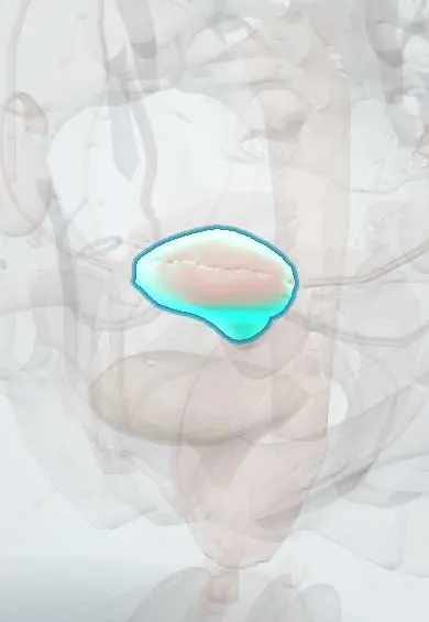
Structures involved
Tap one to see what it is and open it in 3D.
In biochemistry
In histology
In embryology
- Meiosis and nondisjunction: where trisomies come from
- Week 2: the bilaminar disc ('the week of twos')
- Trophoblast and hCG; gestational trophoblastic disease
In physiology
Why it happens
What it is
An abnormal pregnancy in which the placenta (trophoblast) grows as swollen, fluid-filled villi due to an abnormal fertilization. It is a form of gestational trophoblastic disease and can progress to a persistent tumour.
Who gets it
An uncommon complication of pregnancy; texts report higher rates in some parts of Asia and Latin America. Most cases are now detected early by ultrasound in the first trimester.
Causes
- Complete mole: an egg without maternal chromosomes fertilized by one or two sperm (diploid, all paternal); no fetus forms
- Partial mole: a normal egg fertilized by two sperm (triploid); fetal parts may be present
- The excess paternal genetic material drives trophoblast overgrowth
Risk factors
- Maternal age under about 20 or over about 35–40
- Previous molar pregnancy
- Previous miscarriages
- Some dietary factors such as low carotene intake have been proposed
Pathophysiology
With too much paternal genetic material, the trophoblast proliferates and the chorionic villi swell into grape-like vesicles. The abnormal tissue produces very high amounts of human chorionic gonadotropin (hCG), which can stimulate the ovaries (theca lutein cysts) and the thyroid. Some trophoblast can persist or invade the myometrium after evacuation (gestational trophoblastic neoplasia).
Classification
- Complete hydatidiform mole: 46,XX or 46,XY, all paternal, no fetal tissue, higher risk of neoplasia
- Partial hydatidiform mole: usually 69 chromosomes (triploid), fetal tissue may be present, lower risk
- Gestational trophoblastic neoplasia after a mole: invasive mole, choriocarcinoma and rarer forms
Symptoms and signs
- Vaginal bleeding in the first trimester, sometimes passing grape-like tissue
- Uterus larger than expected for the dates (more often in complete mole)
- Severe nausea and vomiting (hyperemesis)
- Absent fetal heart in complete mole
- Less often: early preeclampsia (before 20 weeks), signs of hyperthyroidism, enlarged ovaries with theca lutein cysts
- Many cases are found on a routine early ultrasound without symptoms
Diagnosis
- Clinical
- Suspected from first-trimester bleeding, a large-for-dates uterus or hyperemesis; confirmed by histology of the evacuated tissue.
- Tests
- Quantitative serum hCG, often markedly elevated; blood count, blood group and Rh, kidney, liver and thyroid function; histopathology (and sometimes ploidy or p57 staining) to separate complete from partial mole.
- Imaging
- Pelvic ultrasound: a complete mole shows a heterogeneous vesicular mass without a fetus ("snowstorm" pattern); partial mole shows a thickened cystic placenta, sometimes with an abnormal fetus. Chest X-ray if neoplasia is suspected.
Differential diagnosis
- Miscarriage (incomplete or missed)
- Ectopic pregnancy
- Multiple pregnancy
- Hyperemesis gravidarum of a normal pregnancy
- Uterine fibroids with pregnancy
Goal of treatment
Remove all molar tissue safely, follow hCG to normal to detect persistent disease early, and preserve fertility when desired.
Treatment
- Surgical
- Suction evacuation of the uterus, regardless of uterine size, with oxytocic drugs given only after evacuation begins if bleeding is heavy; hysterectomy is an option for women who do not want more children.
- Medical
- Anti-D immunoglobulin for Rh-negative women; treatment of hyperthyroidism, preeclampsia and anaemia when present.
- Follow-up
- Serial hCG measurements until normal and then for a defined period by protocol, with reliable hormonal contraception during follow-up to avoid confusion with a new pregnancy.
- Medical
- Chemotherapy (single-agent antimetabolite or multi-agent regimens, by risk score) for gestational trophoblastic neoplasia.
Complications
- Haemorrhage and anaemia
- Uterine perforation during evacuation
- Gestational trophoblastic neoplasia: invasive mole, choriocarcinoma
- Hyperthyroidism and, rarely, thyroid storm
- Early preeclampsia
- Respiratory distress from trophoblastic embolization
- Theca lutein cysts with torsion or rupture
Prognosis
Excellent with evacuation and complete hCG follow-up; even trophoblastic neoplasia is highly curable with chemotherapy. The risk of another mole in a future pregnancy is higher than average, so early ultrasound is advised in later pregnancies.
Test yourself
Think of the answer, then tap the question to see it.
What is the karyotype of a complete hydatidiform mole?
Usually 46,XX (sometimes 46,XY), with all chromosomes of paternal origin and no fetal tissue.
How does a partial mole differ genetically from a complete mole?
A partial mole is usually triploid (69 chromosomes, two paternal sets and one maternal) and may contain fetal parts.
Which tumour marker is used to monitor a molar pregnancy?
Serum human chorionic gonadotropin (hCG), followed serially until it normalizes.
What is the classic ultrasound appearance of a complete mole?
A heterogeneous vesicular intrauterine mass without a fetus, described as a snowstorm pattern.
Why can a molar pregnancy cause hyperthyroidism?
Very high hCG shares a subunit with TSH and stimulates the thyroid TSH receptor.
Related conditions
- Miscarriage (early pregnancy loss)
- Ectopic pregnancy
- Preeclampsia
- Ovarian cyst
- Uterine fibroids (leiomyomas)
Educational information, not medical advice. For any symptom, see a health professional.
Sources: Hydatidiform Mole (StatPearls (NCBI Bookshelf), 2024).
Spotted a mistake?We are always improving for the community. Tell us what is wrong and our team will review it.Report a mistake
Representative model, for educational purposes only.