Parapneumonic effusion and empyema !
A parapneumonic effusion is fluid around the lung during pneumonia; when it becomes infected or turns to pus it is called empyema and needs drainage.
Updated 2026-11-01
Structures involved
Tap one to see what it is and open it in 3D.
- Pleura
- Right lower lobe
- Left lower lobe
- Ribs (back part)
- Intercostal nerves
- Diaphragm
- External intercostals
In the clinical exam
Why it happens
- Patterns of acute inflammation: serous, fibrinous, purulent, abscess, ulcer
- Outcomes of acute inflammation: resolution, abscess, scar or chronic inflammation
- Pleural fluid: formation and drainage
- Serous membranes: pleura and peritoneum
What it is
A pleural effusion that develops next to a bacterial pneumonia or lung abscess. It may be simple (sterile exudate), complicated (bacteria invade the pleural space, pH and glucose fall) or frank empyema (pus in the pleural space).
Who gets it
Parapneumonic effusions appear in a large share of patients admitted with bacterial pneumonia, and a small fraction progress to empyema. Empyema is more common in children, older adults, people with alcohol use disorder, diabetes or poor dental health. In Colombia and Latin America tuberculous pleural effusion is an important alternative cause of exudate that must be considered.
Causes
- Bacterial pneumonia (Streptococcus pneumoniae, Staphylococcus aureus, Streptococcus anginosus group)
- Anaerobic and mixed oral flora after aspiration or with a lung abscess
- Gram-negative bacilli in hospital-acquired pneumonia
- Chest trauma, thoracic surgery or pleural procedures
- Spread from a subphrenic abscess or oesophageal rupture
Risk factors
- Delayed treatment of pneumonia
- Aspiration risk: alcohol use disorder, reduced consciousness, dysphagia
- Diabetes mellitus and immunosuppression
- Poor dental hygiene
- Older age and chronic lung disease
Pathophysiology
Develops in three stages. In the exudative stage, inflammation makes the pleura leaky and sterile fluid accumulates. In the fibrinopurulent stage, bacteria invade, neutrophils and fibrin accumulate, pleural fluid pH and glucose fall and loculations form. In the organising stage, fibroblasts create a thick pleural peel that traps the lung and limits its expansion.
Classification
- Simple (uncomplicated) parapneumonic effusion: free-flowing, sterile, pH 7.20 or higher
- Complicated parapneumonic effusion: pH below 7.20, low glucose, high LDH or positive Gram stain or culture
- Empyema: frank pus in the pleural space
- Stages: exudative, fibrinopurulent, organising
Symptoms and signs
- Fever persisting or returning despite treatment of pneumonia
- Pleuritic chest pain that worsens with breathing
- Shortness of breath and cough
- Dullness to percussion, reduced breath sounds and reduced tactile fremitus on the affected side
- Night sweats, weight loss and malaise in subacute cases
Diagnosis
- Clinical
- Suspected in a patient with pneumonia whose fever or pain persists, with signs of an effusion on examination. Every effusion of significant size in a patient with pneumonia is sampled.
- Tests
- Diagnostic thoracentesis is the key test: appearance, pH, glucose, LDH, protein, cell count, Gram stain and culture; Light criteria confirm an exudate. Pus, pH below 7.20 or a positive Gram stain or culture indicate the need for drainage. Blood cultures, blood count and C-reactive protein; adenosine deaminase and mycobacterial testing when tuberculosis is possible.
- Imaging
- Chest X-ray (blunted costophrenic angle, meniscus sign); pleural ultrasound to detect small effusions, septations and to guide the needle; contrast CT shows thickened, enhancing pleura (split pleura sign) and loculations.
Differential diagnosis
- Tuberculous pleural effusion
- Malignant pleural effusion
- Heart failure transudate
- Pulmonary embolism with effusion
- Lung abscess with air-fluid level
Goal of treatment
Control the infection, drain the pleural space completely and allow the lung to re-expand before a fibrous peel forms.
Treatment
- Medical
- Prolonged intravenous then oral antibiotics covering the likely organisms, including anaerobes in community-acquired cases (e.g. a beta-lactam with beta-lactamase inhibitor).
- Procedure
- Chest tube drainage for empyema or complicated effusion, usually ultrasound-guided; intrapleural fibrinolytic plus DNase therapy for loculated collections.
- Surgical
- Video-assisted thoracoscopic surgery (VATS) debridement, or open decortication for an organised peel that traps the lung.
- Follow-up
- Nursing care: chest drain function and output, temperature curve, oxygen saturation, pain control and early mobilisation with breathing exercises.
Complications
- Trapped lung and fibrothorax with restrictive defect
- Bronchopleural fistula
- Empyema necessitans (pus tracking through the chest wall)
- Sepsis and death
- Prolonged hospital stay
Prognosis
Most patients recover fully with antibiotics and adequate drainage. Mortality rises with older age, comorbidity, hospital-acquired infection and delayed drainage.
Prevention
- Early diagnosis and treatment of pneumonia
- Pneumococcal and influenza vaccination
- Good dental hygiene and measures that reduce aspiration
Test yourself
Think of the answer, then tap the question to see it.
Which pleural fluid pH suggests a complicated parapneumonic effusion that needs drainage?
A pH below 7.20, especially with low glucose, high LDH or a positive Gram stain or culture.
What defines empyema?
The presence of frank pus, or organisms on Gram stain, in the pleural space.
What are the three stages of empyema?
Exudative, fibrinopurulent (bacteria, fibrin, loculations) and organising (fibrous peel that traps the lung).
Besides antibiotics, what is the cornerstone of empyema treatment?
Drainage of the pleural space, usually with a chest tube; thoracoscopic surgery or decortication if drainage fails or a peel forms.
Related conditions
- Community-acquired pneumonia
- Pleural effusion
- Lung abscess
- Aspiration pneumonia and pneumonitis
- Hospital-acquired and ventilator-associated pneumonia
- Sepsis and septic shock
- Pulmonary tuberculosis
- Pneumothorax
- Atelectasis
Educational information, not medical advice. For any symptom, see a health professional.
Sources: Empyema (MedlinePlus Medical Encyclopedia (U.S. National Library of Medicine), 2024).
Spotted a mistake?We are always improving for the community. Tell us what is wrong and our team will review it.Report a mistake
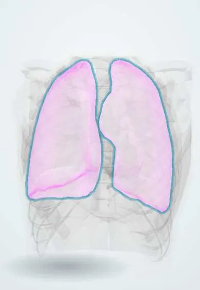
Representative model, for educational purposes only.