Pneumothorax !
A pneumothorax is air trapped between the lung and the chest wall that partly or fully collapses the lung; the tension type is life-threatening.
Updated 2026-10-29
Structures involved
Tap one to see what it is and open it in 3D.
- Pleura
- Right upper lobe
- Left upper lobe
- Ribs (back part)
- Intercostal nerves
- Posterior intercostal arteries
- Superior vena cava
- Trachea
In histology
In the clinical exam
- Chest palpation: expansion and tactile fremitus
- Chest percussion: resonance, dullness and hyperresonance
- The pleuropulmonary syndromes: the comparison table to know
- Pneumothorax syndrome
- Respiratory distress and respiratory failure: the signs
- Pulmonary hyperinflation syndrome (emphysema)
In physiology
- Diving, decompression and hyperbaric oxygen
- Aviation and space physiology
- Mechanics of breathing: pressures and muscles
- Pleural fluid: formation and drainage
Why it happens
What it is
Air in the pleural space that partly or fully collapses the lung. It can be spontaneous, traumatic or iatrogenic; tension pneumothorax is a life-threatening emergency.
Who gets it
Primary spontaneous pneumothorax typically affects tall, thin young men, many of them smokers. Secondary forms occur in older people with lung disease such as COPD or tuberculosis; traumatic pneumothorax is common after chest injury.
Causes
- Primary spontaneous: rupture of small apical blebs, no known lung disease
- Secondary spontaneous: COPD, tuberculosis, pneumonia, cystic lung disease
- Traumatic: penetrating or blunt chest injury, rib fractures
- Iatrogenic: central line insertion, lung biopsy, mechanical ventilation
Risk factors
- Tall, thin young male
- Smoking (tobacco and cannabis)
- Underlying lung disease
- Previous pneumothorax
- Positive-pressure ventilation
- Family history (rare connective tissue or genetic conditions)
Pathophysiology
Air enters the pleural space and the lung's elastic recoil makes it collapse. In tension pneumothorax a one-way valve lets air in but not out, so pressure rises, shifts the mediastinum, kinks the great veins and cuts venous return, causing obstructive shock.
Classification
- Primary spontaneous
- Secondary spontaneous
- Traumatic (open or closed)
- Iatrogenic
- Tension pneumothorax (any cause)
Symptoms and signs
- Sudden one-sided pleuritic chest pain
- Shortness of breath
- Reduced breath sounds on the affected side
- Hyper-resonant percussion
- Reduced chest expansion
- Tension: distress, tachycardia, hypotension, distended neck veins, tracheal deviation away from the affected side
Diagnosis
- Clinical
- Sudden pleuritic pain and breathlessness with reduced breath sounds. Tension pneumothorax is a clinical diagnosis: it is treated immediately, without waiting for imaging.
- Tests
- Pulse oximetry and blood gases assess oxygenation; ECG to exclude cardiac causes of chest pain.
- Imaging
- Chest X-ray shows a visceral pleural line without lung markings beyond it. Bedside ultrasound (absent lung sliding) is fast and sensitive; CT for small or complex cases.
Differential diagnosis
- Pulmonary embolism
- Acute myocardial infarction
- Pneumonia
- Large bulla in emphysema
- Cardiac tamponade (in trauma)
Goal of treatment
Relieve tension immediately when present, re-expand the lung, relieve symptoms and prevent recurrence.
Treatment
- Acute
- Tension pneumothorax: immediate needle or finger decompression followed by a chest drain.
- Conservative
- Small primary spontaneous pneumothorax with few symptoms: observation with oxygen and follow-up imaging.
- Procedure
- Needle aspiration for larger or symptomatic primary pneumothorax, according to guidelines.
- Procedure
- Chest drain (tube thoracostomy) for secondary, traumatic or recurrent cases and for patients on ventilation.
- Surgical
- Video-assisted thoracoscopic surgery with bleb resection and pleurodesis for persistent air leak or recurrence.
Complications
- Tension pneumothorax and cardiac arrest
- Persistent air leak
- Recurrence
- Re-expansion pulmonary oedema
- Haemopneumothorax
Prognosis
Primary spontaneous pneumothorax usually resolves well, but recurrence is common, especially in those who keep smoking. Secondary pneumothorax carries higher risk because lung reserve is low.
Prevention
- Smoking cessation
- Pleurodesis or surgery after recurrence in selected patients
- Careful technique during central line insertion and ventilation
Test yourself
Think of the answer, then tap the question to see it.
Who typically develops a primary spontaneous pneumothorax?
Tall, thin young men, often smokers, from rupture of small apical blebs without known lung disease.
Why must tension pneumothorax be treated before imaging?
It is a clinical diagnosis of obstructive shock; delay to get an X-ray can be fatal, so immediate decompression is done.
Which way does the trachea deviate in tension pneumothorax?
Away from the affected side, because rising pleural pressure pushes the mediastinum to the opposite side.
What ultrasound sign suggests pneumothorax?
Absent lung sliding at the pleural line; a lung point, where sliding reappears, confirms it.
Related conditions
- Chronic obstructive pulmonary disease (COPD)
- Pleural effusion
- Pulmonary embolism
- Cardiac tamponade
- Acute pericarditis
- Rib fractures
- Flail chest
- Haemothorax
Educational information, not medical advice. For any symptom, see a health professional.
Sources: Acute Pneumothorax Evaluation and Treatment (StatPearls (NCBI Bookshelf), 2025).
Spotted a mistake?We are always improving for the community. Tell us what is wrong and our team will review it.Report a mistake
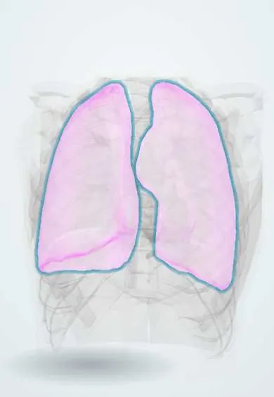
Representative model, for educational purposes only.